Search
- Page Path
- HOME > Search
- Comparison of light-transmittance in dental tissues and dental composite restorations using incremental layering build-up with varying enamel resin layer thickness
- Rodrigo Rocha Maia, Dayane Oliveira, Tracy D'Antonio, Fang Qian, Frederick Skiff
- Restor Dent Endod 2018;43(2):e22. Published online April 16, 2018
- DOI: https://doi.org/10.5395/rde.2018.43.e22

-
 Abstract
 PDF
PDF PubReader
 ePub
ePub Objectives To evaluate and compare light-transmittance in dental tissues and dental composite restorations using the incremental double-layer technique with varying layer thickness.
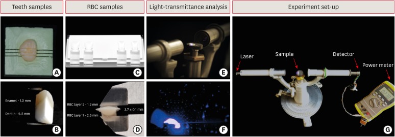
Materials and Methods B1-colored natural teeth slabs were compared to dental restoration build-ups with A2D and B1E-colored nanofilled, supra-nanofilled, microfilled, and microhybrid composites. The enamel layer varied from 0.3, 0.5, or 1.2 mm thick, and the dentin layer was varied to provide a standardized 3.7 mm overall sample thickness (
n = 10). All increments were light-cured to 16 J/cm2 with a multi-wave LED (Valo, Ultradent). Using a spectrophotometer, the samples were irradiated by an RGB laser beam. A voltmeter recorded the light output signal to calculate the light-transmittance through the specimens. The data were analyzed using 1-way analysis of variance followed by thepost hoc Tukey's test (p = 0.05).Results Mean light-transmittance observed at thicker final layers of enamel were significantly lower than those observed at thinner final layers. Within 1.2 mm final enamel resin layer (FERL) thickness, all composites were similar to the dental tissues, with exception of the nanofilled composite. However, within 0.5 mm FERL thickness, only the supra-nanofilled composite showed no difference from the dental tissues. Within 0.3 mm FERL thickness, none of the composites were similar to the dental tissues.
Conclusions The supra-nanofilled composite had the most similar light-transmittance pattern when compared to the natural teeth. However, for other composites, thicker FERL have a greater chance to match the light-transmittance of natural dental tissues.
-
Citations
Citations to this article as recorded by- 3-year randomized clinical trial to evaluate the performance of posterior composite restorations lined with ion-releasing materials
Basma Ahmed, Ramy Ahmed Wafaie, Hamdi H. Hamama, Salah Hasab Mahmoud
Scientific Reports.2024;[Epub] CrossRef - Investigation on the Biaxial Flexural Strength of Universal Shade Resin-Based Composites
Keiko Sakuma, Taku Horie, Takafumi Kishimoto, Mayumi Maesako, Shigetaka Tomoda, Morioki Fujitani, Akimasa Tsujimoto
Polymers.2024; 16(13): 1853. CrossRef - Fabrication of color-graded feldspathic dental prosthetics for aesthetic and restorative dentistry
Imam Akbar Sutejo, Jeehwan Kim, Sinuo Zhang, Chang Woo Gal, Yeong-Jin Choi, Honghyun Park, Hui-suk Yun
Dental Materials.2023; 39(6): 568. CrossRef - Spectrophotometric evaluation of restorative composite shades and their match with a classical shade guide
Rafael Melara, Luciana Mendonça, Fábio Herrmann Coelho-de-Souza, Juliana Nunes Rolla, Luciano de Souza Gonçalves
Restorative Dentistry & Endodontics.2021;[Epub] CrossRef - In vitro wear of dual‐cured bulkfill composites and flowable bulkfill composites
Jean‐François Roulet, Snigdha Gummadi, Hind S. Hussein, Nader Abdulhameed, Chiayi Shen
Journal of Esthetic and Restorative Dentistry.2020; 32(5): 512. CrossRef
- 3-year randomized clinical trial to evaluate the performance of posterior composite restorations lined with ion-releasing materials
- 1,495 View
- 14 Download
- 5 Crossref

- Comparison of the residual stress of the nanofilled composites
- Jeong-won Park
- J Korean Acad Conserv Dent 2008;33(5):457-462. Published online September 30, 2008
- DOI: https://doi.org/10.5395/JKACD.2008.33.5.457
-
 Abstract
 PDF
PDF PubReader
 ePub
ePub "Residual stress" can be developed during polymerization of the dental composite and it can be remained after this process was completed. The total amount of the force which applied to the composite restoration can be calculated by the sum of external and internal force. For the complete understanding of the restoration failure behavior, these two factors should be considered. In this experiment, I compared the residual stress of the recently developed nanofilled dental composite by ring slitting methods.
The composites used in this study can be categorized in two groups, one is microhybrid type-Z250, as control group, and nanofilled type-Grandio, Filtek Supreme, Ceram-X, as experimental ones. Composite ring was made and marked two reference points on the surface. Then measure the change of the distance between these two points before and after ring slitting. From the distance change, average circumferential residual stress (σθ) was calculated. In 10 minutes and 1 hour measurement groups, Filtek Supreme showed higher residual stress than Z250 and Ceram-X. In 24 hour group, Filtek showed higher stress than the other groups.
Following the result of this experiment, nanofilled composite showed similar or higher residual stress than Z250, and when comparing the Z250 and Filtek Supreme, which have quite similar matrix components, Filtek Supreme groups showed higher residual stress.
-
Citations
Citations to this article as recorded by- Microleakage of the experimental composite resin with three component photoinitiator systems
Ji-Hoon Kim, Dong-Hoon Shin
Journal of Korean Academy of Conservative Dentistry.2009; 34(4): 333. CrossRef
- Microleakage of the experimental composite resin with three component photoinitiator systems
- 1,134 View
- 4 Download
- 1 Crossref

- Surface roughness and color stability of various composite resins
- Sung-Yi Lee, Hyeon-Cheol Kim, Bock Hur, Jeong-Kil Park
- J Korean Acad Conserv Dent 2007;32(6):542-549. Published online November 30, 2007
- DOI: https://doi.org/10.5395/JKACD.2007.32.6.542
-
 Abstract
 PDF
PDF PubReader
 ePub
ePub The purpose of this study was to evaluate the difference in the surface roughness after polishing and to evaluate the difference in color stability after immersion in a dye solution among four types of composite resin materials. Four light-polymerized composite resins (Shade A2) with different sized filler content (a nanofilled, a hybrid, a microfilled, a flowble) were used. Average surface roughness (Ra) was measured with a surface roughness tester (Surftest Formtracer) before and after polishing with aluminum oxide abrasive discs (Super-Snap). Color of specimens before and after staining with 2% methylene blue solution were measured using spectrophotometer (CM-3700d) with SCI geometries. The results of Ra and ΔE were analyzed by one-way analysis of variance (ANOVA), a Scheffe multiple comparison test and Student t-test (p = 0.05). After polishing, Ra values were decreased regardless of type of composite resins. In surface roughness after polishing and color stability after staining, nanofilled composite resin was not different with other composite resins except flowable resins.
-
Citations
Citations to this article as recorded by- Effect of contemporary polishing systems on hardness and roughness of one-shaded dental composites
Kivanc Dulger, Gencaga Purcek
Journal of the Australian Ceramic Society.2025; 61(3): 841. CrossRef - Physicomechanical properties and polymerization shrinkage of the newly developed radiopaque flowable composite derived from rice husk
Nor Ain Fatihah Azlisham, Yanti Johari, Dasmawati Mohamad, Mohd Firdaus Yhaya, Zuliani Mahmood
Polymer Composites.2025; 46(7): 5924. CrossRef - Highly Filled Flowable Composite Resins as Sole Restorative Materials: A Systematic Review
Konstantinos Tzimas, Eftychia Pappa, Maria Fostiropoulou, Efstratios Papazoglou, Christos Rahiotis
Materials.2025; 18(14): 3370. CrossRef - Effect of immersion and thermocycling in different beverages on the surface roughness of single- and multi-shade resin composites
Aiah A. El-Rashidy, Omar Shaalan, Rasha M. Abdelraouf, Nour A. Habib
BMC Oral Health.2023;[Epub] CrossRef - Degree of conversion and physicomechanical properties of newly developed flowable composite derived from rice husk using urethane dimethacrylate monomer
Nor Ain Fatihah Azlisham, Yanti Johari, Dasmawati Mohamad, Mohd Firdaus Yhaya, Zuliani Mahmood
Proceedings of the Institution of Mechanical Engineers, Part H: Journal of Engineering in Medicine.2023; 237(12): 1339. CrossRef - Translucency and Color Stability of a Simplified Shade Nanohybrid Composite after Ultrasonic Scaling and Air-Powder Polishing
Ksenia Babina, Maria Polyakova, Inna Sokhova, Vladlena Doroshina, Alexandr Zaytsev, Elena E. Nikonova, Gleb S. Budylin, Evgeny A. Shirshin, Christian Tantardini, Nina Novozhilova
Nanomaterials.2022; 12(24): 4465. CrossRef - Surface properties and color stability of dental flowable composites influenced by simulated toothbrushing
Guangyun LAI, Liya ZHAO, Jun WANG, Karl-Heinz KUNZELMANN
Dental Materials Journal.2018; 37(5): 717. CrossRef - Topography and surface roughness of fluid resins used as bioprotectors of mini-implants
Rogério Lacerda-Santos, Mirella de Fátima Liberato de Moura, Fabíola Galbiatti Carvalho, Hugo Lemes Carlo, Matheus Melo Pithon, Bruno Alessandro Silva Guedes de Lima, Tibério Andrade dos Passos
Applied Adhesion Science.2014;[Epub] CrossRef
- Effect of contemporary polishing systems on hardness and roughness of one-shaded dental composites
- 2,253 View
- 12 Download
- 8 Crossref

- Effect of intermediate resin hydrophilicity on bond strength of single step adhesive
- Yong-Sung Kim, Sang-Hyuk Park, Gi-Woon Choi, Kyoung-Kyu Choi
- J Korean Acad Conserv Dent 2007;32(5):445-458. Published online September 30, 2007
- DOI: https://doi.org/10.5395/JKACD.2007.32.5.445
-
 Abstract
 PDF
PDF PubReader
 ePub
ePub The purpose of this study was to evaluate the bond strength of a new Single step system with different curing mode composites, and to evaluate the effect of the intermediate resins which have different hydrophilicity on bonding ability by means of the micro shear bond testing and TEM examination for the adhesive interface. The adhesive used in this study was an experimental single step system (Bisco Inc., Schaumburg, IL). Experimental groups were produced by using six kinds of intermediate resin having different hydrophilicity that was hydrophilic, hydrophobic and most hydrophobic resin and as filled or not after applying adhesive. Each experimental group was further divided into two subgroups whether the adhesive was light cured or not. Dual cured composite (Bis Core, Bisco Ltd., Schaumburg, IL) was placed on the adhesive layer as light cure or self cure mode. The results of bond strength were statistically analyzed using one way ANOVA and multiple comparisons are made using Tukey's test at α < 0.05 level.
The results of this study were as follows;
1. The application of intermediate resin did not increase the bond strength for light cured composite.
2. The bond strength of an experimental adhesive with self cured composite was significantly increased by the application of intermediate resin layer.
3. The bond strength of adhesive was irrespective of the cure or not of itself before intermediate resin layer applied.
4. As applied hydrophilic resin layer was, the initial bond strength was higher than both hydrophobic and most hydrophobic one used but there was no significance.
Using a single step adhesive with dual/self cured composite, the incompatibility between both of them should be solved by the application of intermediate hydrophobic resin to reduce the adhesive permeability. However, Single step adhesive can be used in the light cured composite restoration without any decrease of the initial bond strength.
-
Citations
Citations to this article as recorded by- The effect of priming etched dentin with solvent on the microtensile bond strength of hydrophobic dentin adhesive
Eun-Sook Park, Ji-Hyun Bae, Jong-Soon Kim, Jae-Hoon Kim, In-Bog Lee, Chang-Keun Kim, Ho-Hyun Son, Byeong-Hoon Cho
Journal of Korean Academy of Conservative Dentistry.2009; 34(1): 42. CrossRef
- The effect of priming etched dentin with solvent on the microtensile bond strength of hydrophobic dentin adhesive
- 990 View
- 1 Download
- 1 Crossref

- The effect of adhesive thickness on microtensile bond strength to the cavity wall
- Hwa-Eon Lee, Hyeon-Cheol Kim, Bock Hur, Jeong-Kil Park
- J Korean Acad Conserv Dent 2007;32(1):9-18. Published online January 31, 2007
- DOI: https://doi.org/10.5395/JKACD.2007.32.1.009
-
 Abstract
 PDF
PDF PubReader
 ePub
ePub The purposes of this study were to examine the variability of adhesive thickness on the different site of the cavity wall when used total-etch system without filler and simplified self-etch system with filler and to evaluate the relationship between variable adhesive thickness and microtensile bond strength to the cavity wall.
A class I cavity in six human molars was prepared to expose all dentinal walls. Three teeth were bonded with a filled adhesive, Clearfil™ SE bond and the other three teeth were bonded with unfilled adhesives, Scotchbond™ Multi Purpose. Morphology and thickness of adhesive layer were examined using fluorescence microscope. Bonding agent thickness was measured at three points along the axial cavity wall, edge of cavity margin
(rim) (hlf) (ang) rim, hlf ang For both bonding agents, adhesive thickness of
ang rim hlf Adhesive thickness of internal angle of the cavity was significantly thicker than that of the cavity margin and the halfway cavity wall for both bonding agents. Microtensile bond strength of the thick adhesive layer at the internal angle of the cavity was higher than that of the thin adhesive layer at the cavity margin and the halfway cavity in the two bonding systems.
-
Citations
Citations to this article as recorded by- Bond strength of a 3-step total-etch bonding system to dentine – An improved approach
H. Hassan Elnadif, W. Palin, M.A. Hadis, B.W. Darvell
Dental Materials.2025; 41(5): 483. CrossRef - Evaluation of bonding effectiveness of a self-etch and an etch-and-rinse adhesive resin to un-treated and Er:Yag laser treated dentin using mini-interfacial fracture toughness test
Marjan Behroozibakhsh, Sotoudeh Davaie, Abbas Monzavi, Tayebeh Abazari, Sima Shahabi, Maryam Pirmoradian
Journal of Adhesion Science and Technology.2019; 33(11): 1201. CrossRef - The effect of Er,Cr:YSGG irradiation on microtensile bond strength of composite resin restoration
Jeong-Hye Son, Hyeon-Cheol Kim, Bock Hur, Jeong-Kil Park
Journal of Korean Academy of Conservative Dentistry.2010; 35(2): 134. CrossRef
- Bond strength of a 3-step total-etch bonding system to dentine – An improved approach
- 1,614 View
- 5 Download
- 3 Crossref


 KACD
KACD

 First
First Prev
Prev


