Search
- Page Path
- HOME > Search
- Traditional and minimally invasive access cavities in endodontics: a literature review
- Ioanna Kapetanaki, Fotis Dimopoulos, Christos Gogos
- Restor Dent Endod 2021;46(3):e46. Published online August 13, 2021
- DOI: https://doi.org/10.5395/rde.2021.46.e46
-
 Abstract
 PDF
PDF PubReader
 ePub
ePub The aim of this review was to evaluate the effects of different access cavity designs on endodontic treatment and tooth prognosis. Two independent reviewers conducted an unrestricted search of the relevant literature contained in the following electronic databases: PubMed, Science Direct, Scopus, Web of Science, and OpenGrey. The electronic search was supplemented by a manual search during the same time period. The reference lists of the articles that advanced to second-round screening were hand-searched to identify additional potential articles. Experts were also contacted in an effort to learn about possible unpublished or ongoing studies. The benefits of minimally invasive access (MIA) cavities are not yet fully supported by research data. There is no evidence that this approach can replace the traditional approach of straight-line access cavities. Guided endodontics is a new method for teeth with pulp canal calcification and apical infection, but there have been no cost-benefit investigations or time studies to verify these personal opinions. Although the purpose of MIA cavities is to reflect clinicians' interest in retaining a greater amount of the dental substance, traditional cavities are the safer method for effective instrument operation and the prevention of iatrogenic complications.
-
Citations
Citations to this article as recorded by- Benefits of Using Magnification in Access Cavity Preparation by Undergraduate Dental Students: A Micro‐Computed Tomography Study
Manal Almaslamani, Okba Mahmoud, Aya Ali, Mawada Abdelmagied
European Journal of Dental Education.2025;[Epub] CrossRef - Effect of access cavity design on canal instrumentation efficiency and fracture resistance in mandibular molars: A cone-beam computed tomography study
Dalia Al-Harith, Rawan Meshal AlOtaibi
Saudi Journal of Oral Sciences.2025; 12(1): 72. CrossRef - Full-coverage Porcelain-fused-to-metal Crown with Guided Access for Future Endodontic Treatment: A Comparative Pilot In Vitro Study
Mohammed Mashyakhy, Hemant Chourasia, Hafiz Adawi, Abdulaziz Abu-Melha, Elham Khudhayr, Rafif Bakri, Taif Kameli, Khalid Moashy, Hitesh Chohan
The Journal of Contemporary Dental Practice.2025; 26(3): 234. CrossRef - Comparative evaluation of root canal morphology in mandibular first premolars with deep radicular grooves using direct vision, dental operating microscope, 2D radiographic visualisation and micro-computed tomography
Mohmed Isaqali Karobari, Hany Mohamed Aly Ahmed, Mohd Fadhli Bin Khamis, Norliza Ibrahim, Tahir Yusuf Noorani, Miriam Fatima Zaccaro Scelza
PLOS One.2025; 20(7): e0329439. CrossRef - Impact of conservative and traditional endodontic accesses on the strength of maxillary zirconia crowns
Carlos A. Jurado, Gustavo Morrice, Mark Antal, Silvia Rojas‐Rueda, Francisco X. Azpiazu‐Flores, Brian R. Morrow, Franklin Garcia‐Godoy, Damian J. Lee
Journal of Prosthodontics.2025;[Epub] CrossRef - A Finite Element Method Study of Stress Distribution in Dental Hard Tissues: Impact of Access Cavity Design and Restoration Material
Mihaela-Roxana Boțilă, Dragos Laurențiu Popa, Răzvan Mercuț, Monica Mihaela Iacov-Crăițoiu, Monica Scrieciu, Sanda Mihaela Popescu, Veronica Mercuț
Bioengineering.2024; 11(9): 878. CrossRef - Impact of Access Cavity Design on Fracture Resistance of Endodontically Treated Maxillary First Premolar: In Vitro
Anju Daniel, Abdul Rahman Saleh, Anas Al-Jadaa, Waad Kheder
Brazilian Dental Journal.2024;[Epub] CrossRef - Management of Traumatized Teeth With Severely Calcified Canals and Minimally Invasive Access Cavity Using the AReneto® System: A Case Report
Pucha Sai Manaswini, Varun Prabhuji, Champa C, Srirekha A, Veena S Pai
Cureus.2024;[Epub] CrossRef - Exploring the Impact of Access Cavity Designs on Canal Orifice Localization and Debris Presence: A Scoping Review
Mario Dioguardi, Davide La Notte, Diego Sovereto, Cristian Quarta, Andrea Ballini, Vito Crincoli, Riccardo Aiuto, Mario Alovisi, Angelo Martella, Lorenzo Lo Muzio
Clinical and Experimental Dental Research.2024;[Epub] CrossRef - The effect of computer aided navigation techniques on the precision of endodontic access cavities: A systematic review and meta-analysis
P. R. Kesharani, S. D. Aggarwal, N. K. Patel, J. A. Patel, D. A. Patil, S. H. Modi
Endodontics Today.2024; 22(3): 244. CrossRef - Minimally Invasive Access Cavity Designs: A Review
Sushmita Rane, Varsha Pandit, Ashwini Gaikwad, Shivani Chavan, Rajlaxmi Patil, Mrunal Shinde
Journal of Pharmacy and Bioallied Sciences.2024; 16(Suppl 3): S1971. CrossRef - Influence of Cavity Designs on Fracture Resistance: Analysis of the Role of Different Access Techniques to the Endodontic Cavity in the Onset of Fractures: Narrative Review
Mario Dioguardi, Davide La Notte, Diego Sovereto, Cristian Quarta, Angelo Martella, Andrea Ballini, Cornelis H. Pameijer
The Scientific World Journal.2024;[Epub] CrossRef - Digital precision meets dentin preservation: PriciGuide™ system for guided access opening
Varun Prabhuji, A. Srirekha, Veena Pai, Archana Srinivasan, S. M. Laxmikanth, Shwetha Shanbhag
Journal of Conservative Dentistry and Endodontics.2024; 27(8): 884. CrossRef - Minimal Invasive Endodontics: A Comprehensive Narrative Review
Jaydip Marvaniya, Kishan Agarwal, Dhaval N Mehta, Nirav Parmar, Ritwik Shyamal , Jenee Patel
Cureus.2022;[Epub] CrossRef
- Benefits of Using Magnification in Access Cavity Preparation by Undergraduate Dental Students: A Micro‐Computed Tomography Study
- 6,723 View
- 183 Download
- 8 Web of Science
- 14 Crossref

- Ten years of minimally invasive access cavities in Endodontics: a bibliometric analysis of the 25 most-cited studies
- Emmanuel João Nogueira Leal Silva, Karem Paula Pinto, Natasha C. Ajuz, Luciana Moura Sassone
- Restor Dent Endod 2021;46(3):e42. Published online July 21, 2021
- DOI: https://doi.org/10.5395/rde.2021.46.e42

-
 Abstract
 PDF
PDF PubReader
 ePub
ePub Objectives This study aimed to analyze the main features of the 25 most-cited articles in minimally invasive access cavities.
Materials and Methods An electronic search was conducted on the Clarivate Analytics' Web of Science ‘All Databases’ to identify the most-cited articles related to this topic. Citation counts were cross-matched with data from Elsevier's Scopus and Google Scholar. Information about authors, contributing institutions and countries, year and journal of publication, study design and topic, access cavity, and keywords were analyzed.
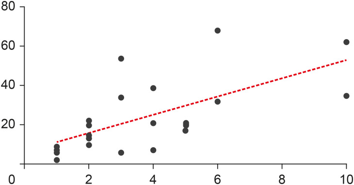
Results The top 25 most-cited articles received a total of 572 (Web of Science), 1,160 (Google Scholar) and 631 (Scopus) citations. It was observed a positive significant association between the number of citations and age of publication (
r = 0.6907,p < 0.0001); however, there was no significant association regarding citation density and age of publication (r = −0.2631,p = 0.2038). TheJournal of Endodontics made the highest contribution (n = 15, 60%). The United States had the largest number of publications (n = 7) followed by Brazil (n = 4), with the most contributions from the University of Tennessee and Grande Rio University (n = 3), respectively. The highest number of most-cited articles wereex vivo studies (n = 16), and ‘fracture resistance’ was the major topic studied (n = 10).Conclusions This study revealed a growing interest for researchers in the field of minimally invasive access cavities. Future trends are focused on the expansion of collaborative networks and the conduction of laboratory studies on under-investigated parameters.
-
Citations
Citations to this article as recorded by- Research Trends in Internal Root Resorption from 1947 to 2022: A Bibliometric Analysis of the 50 Most-cited Articles
Laise Pena Braga Monteiro, Larissa Pillar Gomes Martel, Roberta Fonseca de Castro, Emmanuel João Nogueira Leal da Silva, Juliana Melo da Silva Brandão
Journal of Advanced Oral Research.2025; 16(2): 196. CrossRef - Bibliometric analysis of the publications that list the most-cited articles in endodontics
Oscar Alejandro Gutiérrez-Alvarez, Luis Alberto Pantoja-Villa, Benigno Miguel Calderón-Rojas
Endodontology.2025; 37(2): 128. CrossRef - Sixty Years of Ethylenediaminetetraacetic Acid Use in Endodontics: A Comprehensive Bibliometric Study
Camila Segatto Hartmann, Luiz Fernando Monteiro Czornobay, Julia Menezes Savaris, Aurélio de Oliveira Rocha, Lucas Menezes dos Anjos, Bruno Alexandre Pacheco de Castro Henriques, Mariane Cardoso, Lucas da Fonseca Roberti Garcia, Cleonice da Silveira Teixe
Journal of Endodontics.2025;[Epub] CrossRef - Evaluation of the forces applied by rubber dam clamps on mandibular first molar teeth with different endodontic access cavities: a 3D FEA study
Mehmet Eskibağlar, Serkan Erdem, Büşra Karaağaç Eskibağlar, Mete Onur Kaman
PeerJ.2024; 12: e17921. CrossRef - A Global Overview of Guided Endodontics: A Bibliometric Analysis
Thaine Oliveira Lima, Aurélio de Oliveira Rocha, Lucas Menezes dos Anjos, Nailson Silva Meneses Júnior, Marco Antonio Hungaro Duarte, Murilo Priori Alcalde, Mariane Cardoso, Rodrigo Ricci Vivan
Journal of Endodontics.2024; 50(1): 10. CrossRef - Novel method for augmented reality guided endodontics: An in vitro study
Marco Farronato, Andres Torres, Mariano S. Pedano, Reinhilde Jacobs
Journal of Dentistry.2023; 132: 104476. CrossRef - Contribution of Türkiye to the Field of Endodontology: A Visualized Bibliometric Analysis Based on Web of Science
Olcay ÖZDEMİR, Yağız ÖZBAY, Neslihan YILMAZ ÇIRAKOĞLU
Medical Records.2023; 5(1): 91. CrossRef - Effect of access cavities on the biomechanics of mandibular molars: a finite element analysis
Xiao Wang, Dan Wang, Yi-rong Wang, Xiao-gang Cheng, Long-xing Ni, Wei Wang, Yu Tian
BMC Oral Health.2023;[Epub] CrossRef - Contemporary research trends on nanoparticles in endodontics: a bibliometric and scientometric analysis of the top 100 most-cited articles
Sıla Nur Usta, Zeliha Uğur-Aydın, Kadriye Demirkaya, Cumhur Aydın
Restorative Dentistry & Endodontics.2023;[Epub] CrossRef - Evolving trend of systematic reviews and meta-analyses in endodontics: A bibliometric study
GalvinSim Siang Lin, JiaZheng Leong, WenXin Chong, MikoChong Kha Chee, ChinSheng Lee, Manahil Maqbool, TahirYusuf Noorani
Saudi Endodontic Journal.2022; 12(3): 236. CrossRef - Global research trends on photodynamic therapy in endodontics: A bibliometric analysis
Lucas Peixoto de Araújo, Wellington Luiz de Oliveira da Rosa, Leandro Bueno Gobbo, Tamares Andrade da Silva, José Flávio Affonso de Almeida, Caio Cezar Randi Ferraz
Photodiagnosis and Photodynamic Therapy.2022; 40: 103039. CrossRef - Minimal Invasive Endodontics: A Comprehensive Narrative Review
Jaydip Marvaniya, Kishan Agarwal, Dhaval N Mehta, Nirav Parmar, Ritwik Shyamal , Jenee Patel
Cureus.2022;[Epub] CrossRef
- Research Trends in Internal Root Resorption from 1947 to 2022: A Bibliometric Analysis of the 50 Most-cited Articles
- 2,928 View
- 28 Download
- 8 Web of Science
- 12 Crossref

- Effects of the endodontic access cavity on apical debris extrusion during root canal preparation using different single-file systems
- Pelin Tüfenkçi, Koray Yılmaz, Mehmet Adigüzel
- Restor Dent Endod 2020;45(3):e33. Published online June 4, 2020
- DOI: https://doi.org/10.5395/rde.2020.45.e33

-
 Abstract
 PDF
PDF PubReader
 ePub
ePub Objectives This study was conducted to evaluate the effects of traditional and contracted endodontic cavity (TEC and CEC) preparation with the use of Reciproc Blue (RPC B) and One Curve (OC) single-file systems on the amount of apical debris extrusion in mandibular first molar root canals.
Materials and Methods Eighty extracted mandibular first molar teeth were randomly assigned to 4 groups (
n = 20) according to the endodontic access cavity shape and the single file system used for root canal preparation (reciprocating motion with the RCP B and rotary motion with the OC): TEC-RPC B, TEC-OC, CEC-RPC B, and CEC-OC. The apically extruded debris during preparation was collected in Eppendorf tubes. The amount of extruded debris was quantified by subtracting the weight of the empty tubes from the weight of the Eppendorf tubes containing the debris. Data were analyzed using 1-way analysis of variance with the Tukeypost hoc test. The level of significance was set atp < 0.05.Results The CEC-RPC B group showed more apical debris extrusion than the TEC-OC and CEC-OC groups (
p < 0.05). There were no statistically significant differences in the amount of apical debris extrusion among the TEC-OC, CEC-OC, and TEC-RPC B groups.Conclusions RPC B caused more apical debris extrusion in the CEC groups than did the OC single-file system. Therefore, it is suggested that the RPC B file should be used carefully in teeth with a CEC.
-
Citations
Citations to this article as recorded by- Comparative Evaluation of Periapical Expulsion Using Manual, Rotary, and Reciprocating Instrumentation With EndoVac Irrigation: An In Vitro Study
Sachin Metkari, Sanpreet S Sachdev, Pravin Patil, Manoj Ramugade, Kishor D Sapkale, Kulvinder S Banga, Dinesh Rao
Cureus.2025;[Epub] CrossRef - Comparison of Debris Extrusion and Preparation Time by Traverse, R‐Motion Glider C, and Other Glide Path Systems in Severely Curved Canals
Taher Al Omari, Layla Hassouneh, Khawlah Albashaireh, Alaa Dkmak, Rami Albanna, Ali Al-Mohammed, Ahmed Jamleh, Lucas da Fonseca Roberti Garcia
International Journal of Dentistry.2025;[Epub] CrossRef - Minimal İnvaziv Giriş Kavitelerinin Alt Kesici Dişlerdeki Apikal Ekstrüzyona Etkisi
İrem Haskarabağ, Cangül Keskin
Türk Diş Hekimliği Araştırma Dergisi.2025; 4(2): 75. CrossRef - Evaluation of apically extruded debris from root canal filling removal of the mesiobuccal canal of maxillary molars using XP shaper and protaper with two different irrigation
Sanaz Mirsattari, Maryam Zare Jahromi, Masoud Khabiri
Dental Research Journal.2024;[Epub] CrossRef - The Impact of Minimum Invasive Access Cavity Design on the Quality of Instrumentation of Root Canals of Maxillary Molars Using Cone-Beam Computed Tomography: An in Vitro Study
Fahad H Baabdullah, Samia M Elsherief , Rayan A Hawsawi, Hetaf S Redwan
Cureus.2024;[Epub] CrossRef - Assessment of Bacterial Load and Post-Endodontic Pain after One-Visit Root Canal Treatment Using Two Types of Endodontic Access Openings: A Randomized Controlled Clinical Trial
Ahmed M. Al-Ani, Ahmed H. Ali, Garrit Koller
Dentistry Journal.2024; 12(4): 88. CrossRef - The effect of different kinematics on apical debris extrusion with a single-file system
Taher M. N. Al Omari, Giusy Rita Maria La Rosa, Rami Haitham Issa Albanna, Abedelmalek Tabnjh, Flavia Papale, Eugenio Pedullà
Odontology.2023; 111(4): 910. CrossRef - The effects of laser and ultrasonic irrigation activation methods on smear and debris removal in traditional and conservative endodontic access cavities
Hüseyin Gündüz, Esin Özlek
Lasers in Medical Science.2023;[Epub] CrossRef - Influence of access cavity design, sodium hypochlorite formulation and XP‐endo Shaper usage on apical debris extrusion – A laboratory investigation
Jerry Jose, Aishuwariya Thamilselvan, Kavalipurapu Venkata Teja, Giampiero Rossi–Fedele
Australian Endodontic Journal.2023; 49(1): 6. CrossRef - Apically extruded debris, canal transportation, and shaping ability of nickel-titanium instruments on contracted endodontic cavities in molar teeth
Qinqin Zhang, Jingyi Gu, Jiadi Shen, Ming Ma, Ying Lv, Xin Wei
Journal of Oral Science.2023; 65(4): 203. CrossRef - Impact of contracted endodontic cavities on instrumentation efficacy—A systematic review
Manan Shroff, Karkala Venkappa Kishan, Nimisha Shah, Purnima Saklecha
Australian Endodontic Journal.2023; 49(1): 202. CrossRef - Present status and future directions – Minimal endodontic access cavities
Emmanuel João Nogueira Leal Silva, Gustavo De‐Deus, Erick Miranda Souza, Felipe Gonçalves Belladonna, Daniele Moreira Cavalcante, Marco Simões‐Carvalho, Marco Aurélio Versiani
International Endodontic Journal.2022; 55(S3): 531. CrossRef - Effect of guided conservative endodontic access and different file kinematics on debris extrusion in mesial root of the mandibular molars: An in vitro study
Sathish Sundar, Aswathi Varghese, KrithikaJ Datta, Velmurugan Natanasabapathy
Journal of Conservative Dentistry.2022; 25(5): 547. CrossRef - A critical analysis of research methods and experimental models to study apical extrusion of debris and irrigants
Jale Tanalp
International Endodontic Journal.2022; 55(S1): 153. CrossRef - Current strategies for conservative endodontic access cavity preparation techniques—systematic review, meta-analysis, and decision-making protocol
Benoit Ballester, Thomas Giraud, Hany Mohamed Aly Ahmed, Mohamed Shady Nabhan, Frédéric Bukiet, Maud Guivarc’h
Clinical Oral Investigations.2021; 25(11): 6027. CrossRef - Extrusion of debris with and without intentional foraminal enlargement – A systematic review and meta‐analysis
Ricardo Machado, Gislayne Vigarani, Tainara Macoppi, Ajinkya Pawar, Stella Maria Glaci Reinke, Ana Cristina Kovalik Gonçalves
Australian Endodontic Journal.2021; 47(3): 741. CrossRef - Apical debris extrusion of single-file systems in curved canals
Ecehan Hazar, Olcay Özdemir, Mustafa Murat Koçak, Baran Can Sağlam, Sibel Koçak
Endodontology.2021; 33(3): 128. CrossRef - Quantitative Evaluation of Apically Extruded Debris in Root Canals prepared by Single-file Reciprocating and Single File Rotary Instrumentation Systems: A Comparative In vitro Study
Sonal Sinha, Konark Singh, Anju Singh, Swati Priya, Avanindra Kumar, Sahil Kawle
Journal of Pharmacy and Bioallied Sciences.2021; 13(Suppl 2): S1398. CrossRef - THE INFLUENCE OF DIFFERENT PECKING DEPTH ON AMOUNT OF APICALLY EXTRUDED DEBRIS DURING ROOT CANAL PREPARATION
Fatih ÇAKICI, Busra UYSAL, Elif Bahar CAKİCİ, Adem GUNAYDIN
Atatürk Üniversitesi Diş Hekimliği Fakültesi Dergisi.2021; : 1. CrossRef
- Comparative Evaluation of Periapical Expulsion Using Manual, Rotary, and Reciprocating Instrumentation With EndoVac Irrigation: An In Vitro Study
- 2,426 View
- 24 Download
- 19 Crossref

- Effect of chlorhexidine application on the bond strength of resin core to axial dentin in endodontic cavity
- Yun-Hee Kim, Dong-Hoon Shin
- Restor Dent Endod 2012;37(4):207-214. Published online November 21, 2012
- DOI: https://doi.org/10.5395/rde.2012.37.4.207
-
 Abstract
 PDF
PDF PubReader
 ePub
ePub Objectives This study evaluated the influence of chlorhexidine (CHX) on the microtensile bonds strength (µTBS) of resin core with two adhesive systems to dentin in endodontic cavities.
Materials and Methods Flat dentinal surfaces in 40 molar endodontic cavities were treated with self-etch adhesive system, Contax (DMG) and total-etch adhesive system, Adper Single Bond 2 (3M ESPE) after the following surface treatments: (1) Priming only (Contax), (2) CHX for 15 sec + rinsing + priming (Contax), (3) Etching with priming (Adper Single Bond 2), (4) Etching + CHX for 15 sec + rinsing + priming (Adper Single Bond 2). Resin composite build-ups were made with LuxaCore (DMG) using a bulk method and polymerized for 40 sec. For each condition, half of specimens were submitted to µTBS after 24 hr storage and half of them were submitted to thermocycling of 10,000 cycles between 5℃ and 55℃ before testing. The data were analyzed using ANOVA and independent
t -test at a significance level of 95%.Results CHX pre-treatment did not affect the bond strength of specimens tested at the immediate testing period, regardless of dentin surface treatments. However, after 10,000 thermocycling, all groups showed reduced bond strength. The amount of reduction was greater in groups without CHX treatments than groups with CHX treatment. These characteristics were the same in both self-etch adhesive system and total-etch adhesive system.
Conclusions 2% CHX application for 15 sec proved to alleviate the decrease of bond strength of dentin bonding systems. No significant difference was shown in µTBS between total-etching system and self-etching system.
-
Citations
Citations to this article as recorded by- Micro Tensile bond strength and microleakage assessment of total-etch and self-etch adhesive bonded to carious affected dentin disinfected with Chlorhexidine, Curcumin, and Malachite green
Zeeshan Qamar, Nishath Sayed Abdul, R Naveen Reddy, Mahesh Shenoy, Saleh Alghufaili, Yousef Alqublan, Ali Barakat
Photodiagnosis and Photodynamic Therapy.2023; 43: 103636. CrossRef - The Classification and Selection of Adhesive Agents; an Overview for the General Dentist
Naji Ziad Arandi
Clinical, Cosmetic and Investigational Dentistry.2023; Volume 15: 165. CrossRef - Influence of chlorhexidine 2% and sodium hypochlorite 5.25% on micro-tensile bond strength of universal adhesive system (G-Premio Bond)
Nafiseh Fazelian, Abbas Rahimi Dashtaki, MohammadAmin Eftekharian, Batool Amiri
Brazilian Journal of Oral Sciences.2022;[Epub] CrossRef - Comparative evaluation of the effects of different methods of post space preparation in primary anterior teeth on the fracture resistance of tooth restorations
Bahman Seraj, Sara Ghadimi, Ebrahim Najafpoor, Fatemeh Abdolalian, razieh khanmohammadi
Journal of Dental Research, Dental Clinics, Dental Prospects.2019; 13(2): 141. CrossRef - Chemical, microbial, and host‐related factors: effects on the integrity of dentin and the dentin–biomaterial interface
Marcela T. Carrilho, Fabiana Piveta, Leo Tjäderhane
Endodontic Topics.2015; 33(1): 50. CrossRef - MMP Inhibitors on Dentin Stability
A.F. Montagner, R. Sarkis-Onofre, T. Pereira-Cenci, M.S. Cenci
Journal of Dental Research.2014; 93(8): 733. CrossRef - Thermal cycling for restorative materials: Does a standardized protocol exist in laboratory testing? A literature review
Anna Lucia Morresi, Maurizio D'Amario, Mario Capogreco, Roberto Gatto, Giuseppe Marzo, Camillo D'Arcangelo, Annalisa Monaco
Journal of the Mechanical Behavior of Biomedical Materials.2014; 29: 295. CrossRef
- Micro Tensile bond strength and microleakage assessment of total-etch and self-etch adhesive bonded to carious affected dentin disinfected with Chlorhexidine, Curcumin, and Malachite green
- 1,263 View
- 3 Download
- 7 Crossref


 KACD
KACD

 First
First Prev
Prev


