
 KACD
KACD
Search
- Page Path
- HOME > Search
Research Articles
- Microbial leakage and marginal adaptation of alkasite restorative material in Class II cavities: an in vitro study
- Kah Hup Chew, Norhayati Yusop, Kasmawati Mokhtar, Hafizah Ibrahim
- Restor Dent Endod 2026;51(3):e31. Published online July 28, 2026
- DOI: https://doi.org/10.5395/rde.2026.51.e31
-
 Abstract
 PDF
PDF - Objectives
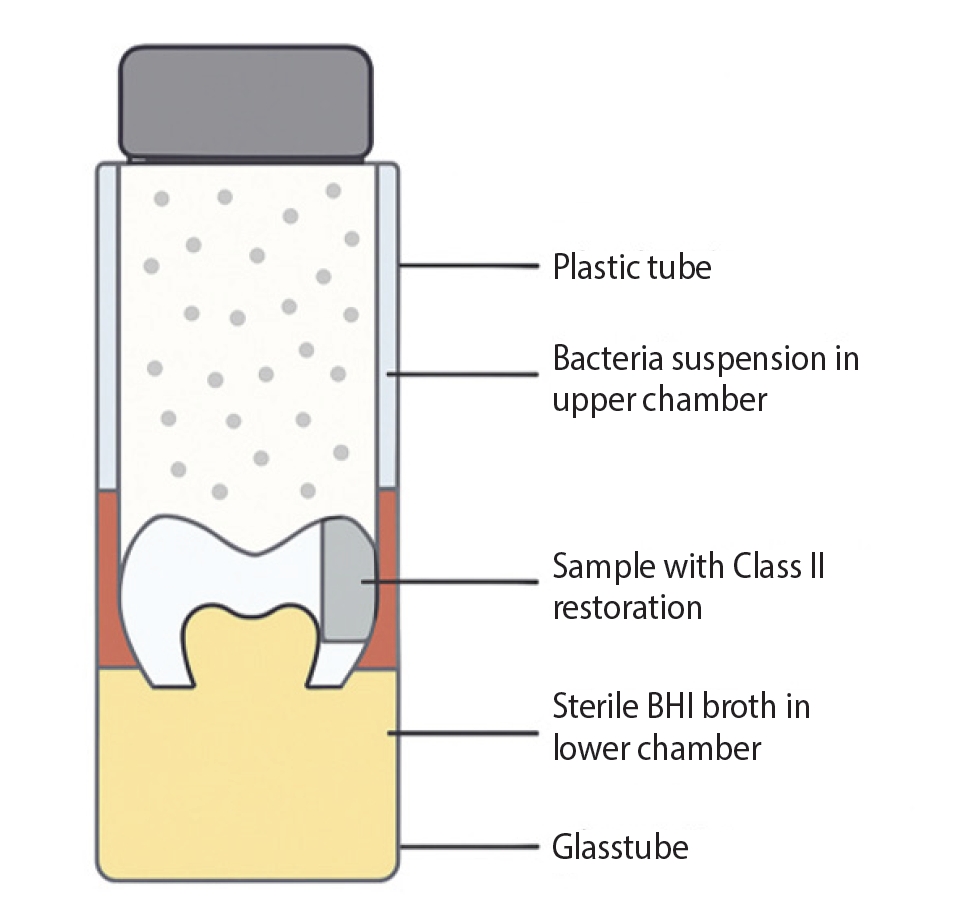
This study aimed to determine the relationship between the interfacial properties of proximal cavities (microbial leakage and marginal adaptation) and restorative materials (alkasite, resin composite, and glass hybrid). Methods: This study involved 66 human third molars that were left intact. Standardized Class II cavities were done on the mesial surface. The samples were then divided into five groups: (i) Cention N (CN, Ivoclar Vivadent), (ii) Filtek Z350XT (FZ, 3M ESPE), (iii) EQUIA Forte (GC Corporation), and (iv) two controls. For 40 days, a dual-chamber model containing Streptococcus mutans was used to study microbial leakage. Qualitative assessments of the sectioned samples were also carried out using a scanning electron microscope (SEM). Moreover, this study employed descriptive and Kaplan-Meier analyses, alongside a one-way analysis of variance for additional descriptive comparisons (p < 0.05). Results: Across different restorative materials, notable variations in microbial leakage were recorded. From the supplementary analysis between CN and FZ, a statistically significant difference was demonstrated (p = 0.037). In comparison to other restorative systems, CN also produced a prolonged mean time before microbial leakage occurred. For the interfacial properties of the restorative systems, illustrative information was offered via the SEM images. Conclusions: Regarding microbial leakage, all restorative systems presented their occurrence (despite the constraints of this study). When assessing CN and FZ restorative systems, a slower rate of microbial leakage was also concluded for CN. For the tooth-restoration interface, qualitative data were verified through SEM observations.
- 1,308 View
- 84 Download

- Marginal adaptation of three root-end filling materials in cavities prepared with laser and ultrasonic tips: an in vitro comparative study
- Busra Zengin, Seda Aydemir, Nicholas Paul Chandler
- Restor Dent Endod 2025;50(4):e32. Published online September 9, 2025
- DOI: https://doi.org/10.5395/rde.2025.50.e32
-
Abstract
PDF
- Objectives
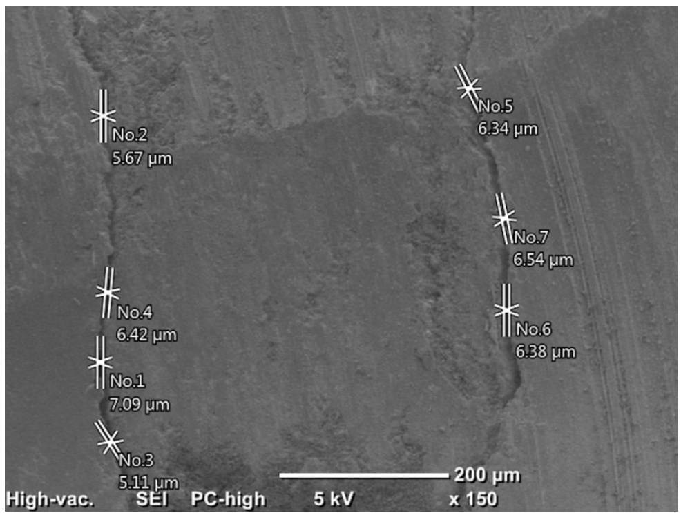
This study evaluated the marginal adaptation of ProRoot MTA (Dentsply Tulsa Dental), Biodentine (Septodont), and TotalFill BC RRM (FKG) placed in root-end cavities prepared with ultrasonic or Er,Cr:YSGG laser tips, using scanning electron microscopy.
Methods
The canals of 90 extracted maxillary central incisors were prepared and obturated and their roots resected. Six groups of 15 specimens were allocated as follows: ultrasonic + ProRoot MTA, ultrasonic + Biodentine, ultrasonic + TotalFill, laser + ProRoot MTA, laser + Biodentine, and laser + TotalFill. Roots were sectioned longitudinally to expose the filling material. Apical and coronal micrographs were taken, and the greatest distance between dentin and filling material was measured. The total gap area was also calculated using further micrographs.
Results
Cavities prepared with the ultrasonic tips and filled with Biodentine showed significantly greater gap dimensions compared with TotalFill (p < 0.001) and ProRoot MTA (p = 0.007) in the apical region. The ultrasonic group showed significantly higher void values compared to the laser group for ProRoot MTA (p = 0.026), when comparing the total values of void. The Biodentine group was significantly higher than the TotalFill group in root-end cavities prepared with ultrasonic tips (p < 0.001). The Biodentine group was significantly higher than the ProRoot MTA group in root-end cavities prepared with the laser tip (p = 0.002).
Conclusions
Under the conditions of this study, it was determined that the root-end cavity preparation technique had an effect on the amount of gaps formed between the dentin and the three filling materials. -
Citations
Citations to this article as recorded by
- Marginal Adaptability of Harvard MTA and Biodentine Used as Root-End Filling Material: A Comparative SEM Study
Yaneta Kouzmanova, Ivanka Dimitrova
Materials.2025; 18(19): 4598. CrossRef
- Marginal Adaptability of Harvard MTA and Biodentine Used as Root-End Filling Material: A Comparative SEM Study
- 4,747 View
- 308 Download
- 1 Web of Science
- 1 Crossref
 First
First Prev
Prev


