
Search
- Page Path
- HOME > Search
Research Articles
- Initial attachment, viability, proliferation, and migration of osteoblast-like SaOS-2 cells on two resorbable xenogeneic membranes for guided tissue regeneration: ab in vitro experimental study
- Rafael Fernández-Grisales, Giovanna García-Suárez, Ximena Guerrero-Rodríguez, Carolina Berruecos-Orozco, Marco Calle-Jaramillo, Wilder Javier Rojas, Vanessa Esmeralda Duque, Daniela Serna-Guisao, Néstor Ríos-Osorio
- Restor Dent Endod 2026;51(2):e20. Published online April 13, 2026
- DOI: https://doi.org/10.5395/rde.2026.51.e20
-
 Abstract
 PDF
PDF PubReader
 ePub
ePub - Objectives
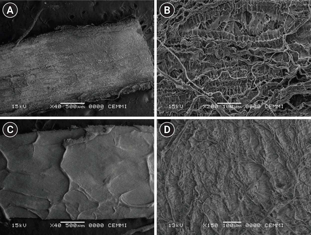
This study evaluated the biocompatibility of a new xenogeneic resorbable membrane derived from porcine esophagus membrane (Quirumatrix, Cells Tech Co.) and compared it with a porcine pericardium membrane (Straumann Jason, Straumann Holding AG.) traditionally used for guided tissue regeneration. The parameters investigated were the viability, migration, and adhesion of SaOS-2 osteoblast-like cells derived from osteosarcoma on both membranes.
Methods
The cells were cultured in 100 mm plates in RPMI 1640 medium (40 mL), supplemented. They were incubated at 37°C in a humidified atmosphere with 95% air and 5% to 10% CO2. Cell morphology and adhesion were evaluated using phase contrast optical microscopy and scanning electron microscope. Cell viability and proliferation were evaluated using a fluorometric resazurin reduction assay, with fluorescence intensity measured at 48, 72, and 96 hours. Cell migration was evaluated using staining with Alexa Fluor 555 Phalloidin (Cell Signaling Technology) and DAPI, with a reference line. Cell migration was analyzed by measuring displacement within the delineated area using an Axio Imager M2 fluorescence microscope (Carl Zeiss). Each membrane was photographed. The statistical analysis was performed using GraphPad Prism ver. 10.2.3 (GraphPad Software). A p-value <0.05 was considered significant between experimental groups.
Results
Both membranes were shown to be biocompatible. The porcine pericardium membrane showed greater cell adhesion and proliferation compared to the porcine esophagus membrane. Cell migration was significantly greater in the Jason membrane.
Conclusions
The results revealed that both evaluated membranes are biocompatible and non-cytotoxic; further research is needed to understand their long-term behavior, interactions with other types of cells, and performance in specific therapeutic situations.
- 1,338 View
- 90 Download

- Antimicrobial and cytotoxic properties of calcium-enriched mixture cement, Iranian propolis, and propolis with herbal extracts in primary dental pulp stem cells
- Mohammad Esmaeilzadeh, Shirin Moradkhani, Fahimeh Daneshyar, Mohammad Reza Arabestani, Sara Soleimani Asl, Soudeh Tayebi, Maryam Farhadian
- Restor Dent Endod 2023;48(1):e2. Published online December 1, 2022
- DOI: https://doi.org/10.5395/rde.2023.48.e2
-
Abstract
PDFPubReaderePub
Objectives In this study, natural substances were introduced as primary dental pulp caps for use in pulp therapy, and the antimicrobial and cytotoxic properties of these substances were investigated.
Materials and Methods In this
in vitro study, the antimicrobial properties of calcium-enriched mixture (CEM) cement, propolis, and propolis individually combined with the extracts of several medicinal plants were investigated againstEnterococcus faecalis ,Escherichia coli ,Pseudomonas aeruginosa , andStaphylococcus aureus . Then, the cytotoxicity of each substance or mixture against pulp stem cells extracted from 30 primary healthy teeth was evaluated at 4 concentrations. Data were gathered via observation, and optical density values were obtained using the 3-(4,5-dimethylthiazol-2-yl)-2,5-diphenyl-2H-tetrazolium bromide (MTT) test and recorded. SPSS software version 23 was used to analyze the data. Data were evaluated using 2-way analysis of variance and the Tukey test.Results Regarding antimicrobial properties, thyme alone and thyme + propolis had the lowest minimum inhibitory concentrations (MICs) against the growth of
S. aureus ,E. coli , andP. aeruginosa bacteria. ForE. faecalis , thyme + propolis had the lowest MIC, followed by thyme alone. At 24 and 72 hours, thyme + propolis, CEM cement, and propolis had the greatest bioviability in the primary dental pulp stem cells, and lavender + propolis had the lowest bioviability.Conclusions Of the studied materials, thyme + propolis showed the best results in the measures of practical performance as a dental pulp cap.
-
Citations
Citations to this article as recorded by
- Self-Adapting Mouthguard with Nano-Hydroxyapatite and Propolis for Early Childhood Caries: Preclinical Safety and Efficacy
Mata Soslanbekovna Mustapaeva, Khadizhat Adamovna Sataeva, Elina Ilyasovna Zhabrailova, Alina Mairbekovna Sidakova, Karina Maharbekovna Mukagova, Umar Said-Asanovich Magomadov, Muslim Usmanovich Dunaev, Mehdi Usmanovich Dunaev, Khava Khuseynovna Amaeva, V
Asian Journal of Periodontics and Orthodontics.2026; 6(1): 32. CrossRef - Comprehensive review of composition, properties, clinical applications, and future perspectives of calcium-enriched mixture (CEM) cement: a systematic analysis
Saeed Asgary, Mahtab Aram, Mahta Fazlyab
BioMedical Engineering OnLine.2024;[Epub] CrossRef - Effects of aqueous and ethanolic extracts of Chinese propolis on dental pulp stem cell viability, migration and cytokine expression
Ha Bin Park, Yen Dinh, Pilar Yesares Rubi, Jennifer L. Gibbs, Benoit Michot
PeerJ.2024; 12: e18742. CrossRef
- Self-Adapting Mouthguard with Nano-Hydroxyapatite and Propolis for Early Childhood Caries: Preclinical Safety and Efficacy
- 3,488 View
- 59 Download
- 2 Web of Science
- 3 Crossref
 First
First Prev
Prev

