Search
- Page Path
- HOME > Search
- A 48-month clinical performance of hybrid ceramic fragment restorations manufactured in CAD/CAM in non-carious cervical lesions: case report
- Michael Willian Favoreto, Gabriel David Cochinski, Eveline Claudia Martini, Thalita de Paris Matos, Matheus Coelho Bandeca, Alessandro Dourado Loguercio
- Restor Dent Endod 2024;49(3):e32. Published online August 5, 2024
- DOI: https://doi.org/10.5395/rde.2024.49.e32

-
 Abstract
 PDF
PDF PubReader
 ePub
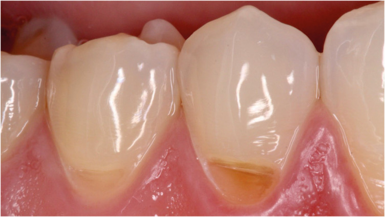
ePub From the restorative perspective, various methods are available to prevent the progression of non-carious cervical lesions. Direct, semi-direct, and indirect composite resin techniques and indirect ceramic restorations are commonly recommended. In this context, semi-direct and indirect restoration approaches are increasingly favored, particularly as digital dentistry becomes more prevalent. To illustrate this, we present a case report demonstrating the efficacy of hybrid ceramic fragments fabricated using computer-aided design (CAD)/computer-aided manufacturing (CAM) technology and cemented with resin cement in treating non-carious cervical lesions over a 48-month follow-up period. A 24-year-old male patient sought treatment for aesthetic concerns and dentin hypersensitivity in the cervical region of the lower premolar teeth. Clinical examination confirmed the presence of two non-carious cervical lesions in the buccal region of teeth #44 and #45. The treatment plan involved indirect restoration using CAD/CAM-fabricated hybrid ceramic fragments as a restorative material. After 48 months, the hybrid ceramic material exhibited excellent adaptation and durability provided by the CAD/CAM system. This case underscores the effectiveness of hybrid ceramic fragments in restoring non-carious cervical lesions, highlighting their long-term stability and clinical success.
- 3,423 View
- 150 Download

- Persistent pain after successful endodontic treatment in a patient with Wegener’s granulomatosis: a case report
- Ricardo Machado, Jorge Aleixo Pereira, Filipe Colombo Vitali, Michele Bolan, Elena Riet Correa Rivero
- Restor Dent Endod 2022;47(3):e26. Published online June 9, 2022
- DOI: https://doi.org/10.5395/rde.2022.47.e26

-
 Abstract
 PDF
PDF PubReader
 ePub
ePub Wegener’s granulomatosis (WG) is a condition with immune-mediated pathogenesis that can present oral manifestations. This report describes the case of a patient diagnosed with WG 14 years previously, who was affected by persistent pain of non-odontogenic origin after successful endodontic treatment. A 39-year-old woman with WG was diagnosed with pulp necrosis and apical periodontitis of teeth #31, #32, and #41, after evaluation through a clinical examination and cone-beam computed tomography (CBCT). At the first appointment, these teeth were subjected to conventional endodontic treatment. At 6- and 12-month follow-up visits, the patient complained of persistent pain associated with the endodontically treated teeth (mainly in tooth #31), despite complete remission of the periapical lesions shown by radiographic and CBCT exams proving the effectiveness of the endodontic treatments, thus indicating a probable diagnostic of persistent pain of non-odontogenic nature. After the surgical procedure was performed to curette the lesion and section 3 mm of the apical third of tooth #31, the histopathological analysis suggested that the painful condition was likely associated with the patient's systemic condition. Based on clinical, radiographic, and histopathological findings, this unusual case report suggests that WG may be related to non-odontogenic persistent pain after successful endodontic treatments.
-
Citations
Citations to this article as recorded by- Toothaches of Non-odontogenic Origin
Davis C. Thomas, Tanvee Somaiya, Ahana Ajayakumar, Vaishnavi Prabhakar
Dental Clinics of North America.2026; 70(1): 209. CrossRef
- Toothaches of Non-odontogenic Origin
- 4,789 View
- 51 Download
- 1 Crossref


 KACD
KACD

 First
First Prev
Prev


