
Previous issues
- Page Path
- HOME > Browse articles > Previous issues
- Volume 50 (4); November 2025
-


Research Articles
- Marginal adaptation of three root-end filling materials in cavities prepared with laser and ultrasonic tips: an in vitro comparative study
- Busra Zengin, Seda Aydemir, Nicholas Paul Chandler
- Restor Dent Endod 2025;50(4):e32. Published online September 9, 2025
- DOI: https://doi.org/10.5395/rde.2025.50.e32
-
 Abstract
 PDF
PDF PubReader
 ePub
ePub - Objectives
This study evaluated the marginal adaptation of ProRoot MTA (Dentsply Tulsa Dental), Biodentine (Septodont), and TotalFill BC RRM (FKG) placed in root-end cavities prepared with ultrasonic or Er,Cr:YSGG laser tips, using scanning electron microscopy.
Methods
The canals of 90 extracted maxillary central incisors were prepared and obturated and their roots resected. Six groups of 15 specimens were allocated as follows: ultrasonic + ProRoot MTA, ultrasonic + Biodentine, ultrasonic + TotalFill, laser + ProRoot MTA, laser + Biodentine, and laser + TotalFill. Roots were sectioned longitudinally to expose the filling material. Apical and coronal micrographs were taken, and the greatest distance between dentin and filling material was measured. The total gap area was also calculated using further micrographs.
Results
Cavities prepared with the ultrasonic tips and filled with Biodentine showed significantly greater gap dimensions compared with TotalFill (p < 0.001) and ProRoot MTA (p = 0.007) in the apical region. The ultrasonic group showed significantly higher void values compared to the laser group for ProRoot MTA (p = 0.026), when comparing the total values of void. The Biodentine group was significantly higher than the TotalFill group in root-end cavities prepared with ultrasonic tips (p < 0.001). The Biodentine group was significantly higher than the ProRoot MTA group in root-end cavities prepared with the laser tip (p = 0.002).
Conclusions
Under the conditions of this study, it was determined that the root-end cavity preparation technique had an effect on the amount of gaps formed between the dentin and the three filling materials. -
Citations
Citations to this article as recorded by
- Marginal Adaptability of Harvard MTA and Biodentine Used as Root-End Filling Material: A Comparative SEM Study
Yaneta Kouzmanova, Ivanka Dimitrova
Materials.2025; 18(19): 4598. CrossRef
- Marginal Adaptability of Harvard MTA and Biodentine Used as Root-End Filling Material: A Comparative SEM Study
- 3,739 View
- 282 Download
- 1 Web of Science
- 1 Crossref

- The influence of bioactive glass (BGS-7) on enamel remineralization: an in vitro study
- Chaeyoung Lee, Eunseon Jeong, Kun-Hwa Sung, Su-Jung Park, Yoorina Choi
- Restor Dent Endod 2025;50(4):e33. Published online October 15, 2025
- DOI: https://doi.org/10.5395/rde.2025.50.e33
-
Abstract
PDFPubReaderePub
- Objectives
The aim of this study was to compare the remineralizing capacity of bioactive glass (BGS-7, CGBIO) with other agents.
Methods
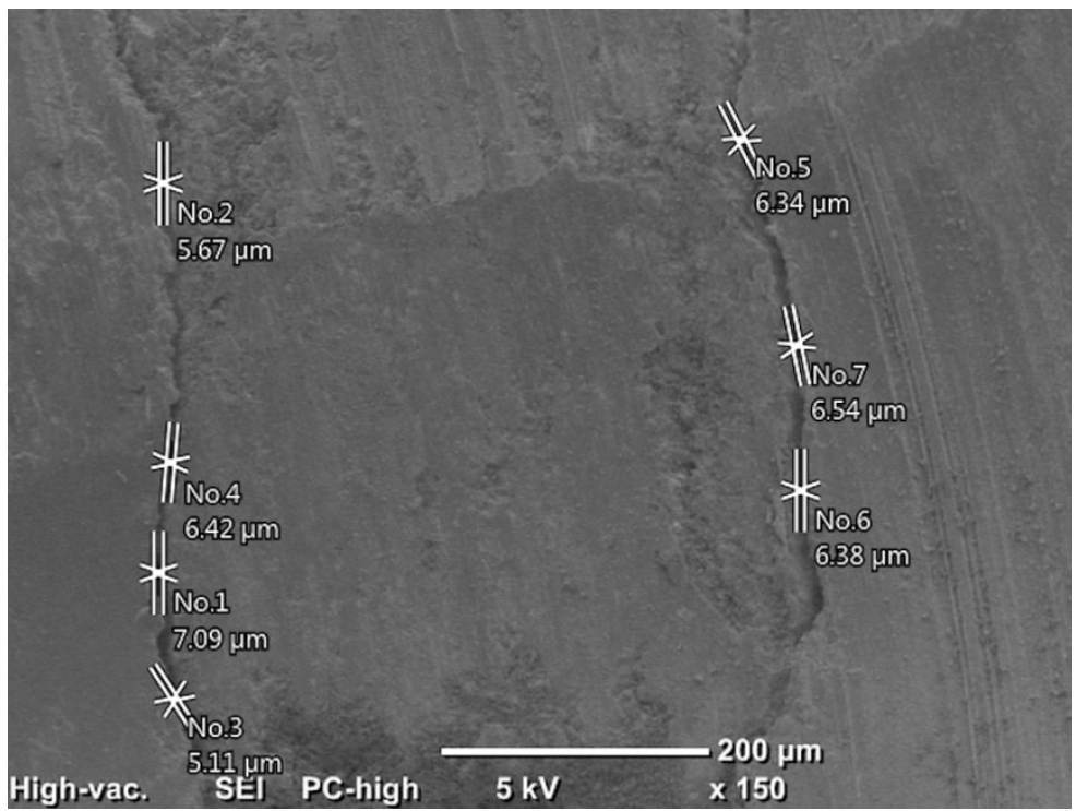
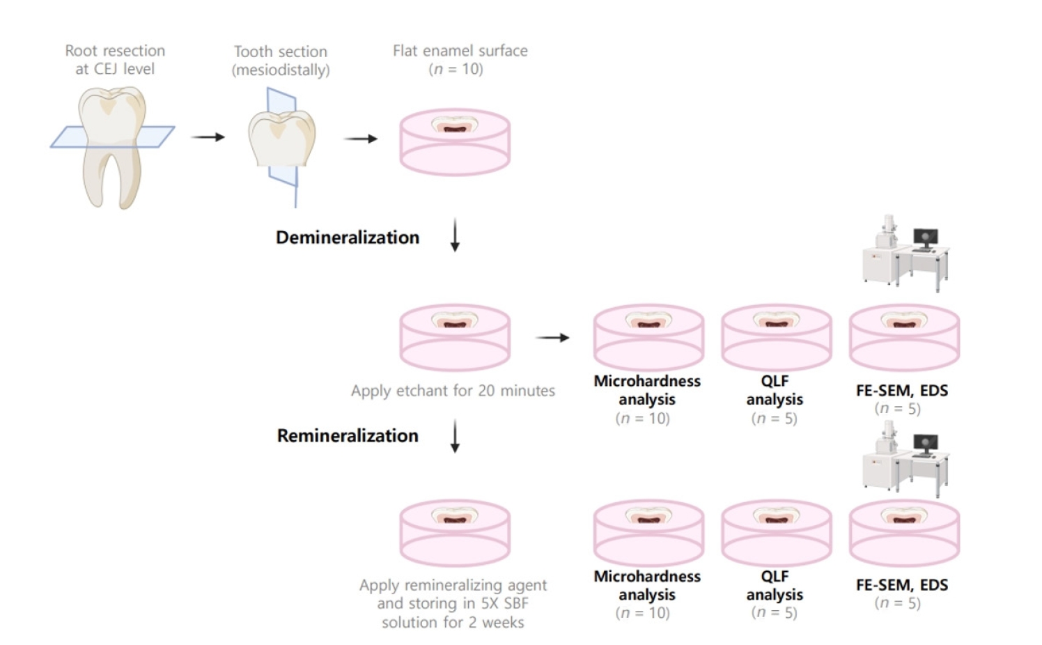
Twenty caries-free third molars were sectioned and demineralized. Specimens were divided into four groups: (1) control, (2) Clinpro XT varnish (Solventum), (3) 1.23% acidulated phosphate fluoride gel, and (4) a new type of CaO-SiO2-P2O5-B2O3 system of bioactive glass ceramics (BGS-7). Agents were applied and stored in simulated body fluid at 37℃ for 2 weeks. Microhardness was measured using the Vickers hardness testing method. Five specimens per group were analyzed using quantitative light-induced fluorescence (QLF) to assess mineral loss. Field-emission scanning electron microscopy (FE-SEM) and energy-dispersive X-ray spectroscopy (EDS) were used to examine the surface morphology and elemental composition. Data were analyzed using paired t-test and one-way analysis of variance (p < 0.05).
Results
BGS-7 showed the highest microhardness values and the greatest recovery in QLF analysis (p < 0.05). FE-SEM revealed granular precipitates on demineralized enamel in the BGS-7 group. EDS confirmed the presence of newly formed silicon and fluoride layers.
Conclusions
BGS-7 demonstrated superior remineralization capacity compared to other agents, suggesting its potential as an effective remineralizing material. -
Citations
Citations to this article as recorded by- Bacterial ghosts (BGs): A promising approach as candidate vaccine
Helal F. Hetta, Ibraheem M. Mwafey, Noura H. Abd Ellah, Fawaz E. Alanazi, Yasmin N. Ramadan
World Journal of Microbiology and Biotechnology.2026;[Epub] CrossRef
- Bacterial ghosts (BGs): A promising approach as candidate vaccine
- 2,431 View
- 228 Download
- 1 Web of Science
- 1 Crossref
- Analysis of temperature change during polymerization according to resin thickness: an in vitro experimental study
- Kkot-Byeol Bae, Eun-Young Noh, Young-Tae Cho, Bin-Na Lee, Hoon-Sang Chang, Yun-Chan Hwang, Won-Mann Oh, In-Nam Hwang
- Restor Dent Endod 2025;50(4):e34. Published online November 12, 2025
- DOI: https://doi.org/10.5395/rde.2025.50.e34
-
Abstract
PDFPubReaderePub
- Objectives
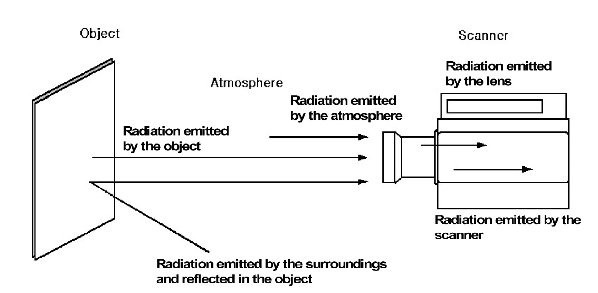
This study aimed to analyze the temperature changes during the light curing of conventional flowable composite resin and bulk-fill composite resin of various thicknesses using an infrared thermographic camera.
Methods
Flowable composite resin (G-aenial Flo, GC Co.) and bulk-fill composite resin (SDR, Dentsply Caulk) were used. Specimens with thicknesses from 0.5 mm to 5.0 mm were prepared. The infrared thermographic camera measured the temperature changes at the maximum temperature rise point during light curing. The data were analyzed for maximum temperature, time to peak temperature, and temperature rise patterns.
Results
For G-aenial Flo, the maximum temperature tended to decrease with increasing thickness, whereas for SDR, the maximum temperature decreased up to 2.0 mm and then remained relatively consistent from 2.0 mm to 5.0 mm. At thicknesses of 1.5 mm or less, both resins showed a rapid temperature increase within the first 5 seconds, followed by a reduced rate of increase up to 80 seconds. At thicknesses of 2.0 mm or greater, the temperature peaked and then gradually decreased. Across all thicknesses, SDR was observed to reach peak temperature more rapidly than G-aenial Flo.
Conclusions
Observable differences in polymerization dynamics were identified between the two resin types, particularly at greater thicknesses. Although no statistical analysis was performed, these descriptive findings suggest that infrared thermographic cameras may be useful for indirectly assessing polymerization dynamics during resin polymerization.
- 1,953 View
- 96 Download
- Phase transformation temperatures influence the reduction ratio of fatigue resistance of nickel-titanium reciprocating files at body temperature: an in vitro experimental study
- Walid Nehme, Alfred Naaman, Lola Pedèches, Sylvie Lê, Marie Georgelin-Gurgel, Sang Won Kwak, Hyeon-Cheol Kim, Franck Diemer
- Restor Dent Endod 2025;50(4):e35. Published online November 5, 2025
- DOI: https://doi.org/10.5395/rde.2025.50.e35
-
Abstract
PDFPubReaderePub
- Objectives
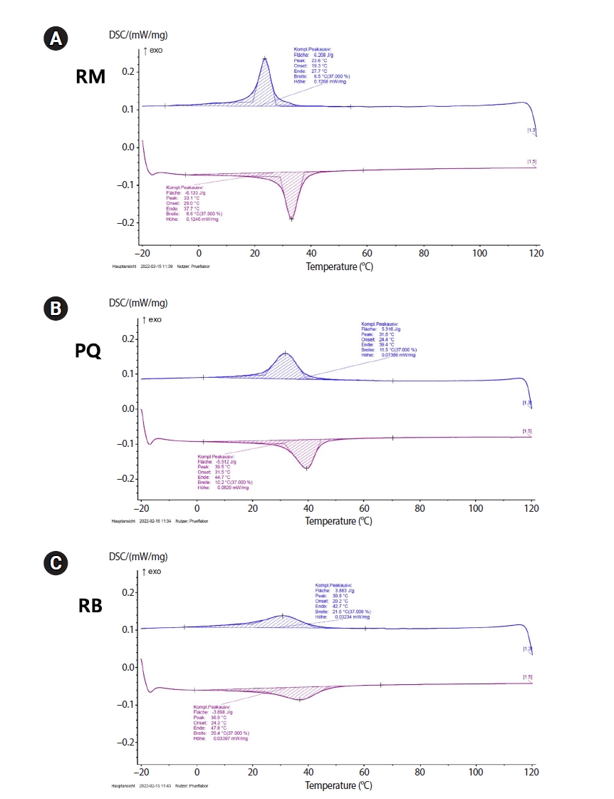
The objective of this study was to evaluate the effects of transformational temperatures on the cyclic fatigue resistance at body temperature of reciprocating file systems: R motion (RM), Procodile Q (PQ), and Reciproc Blue.
Methods
Resistance test was done in a custom-made device at room (20°C ± 1°C) and body (37°C ± 1°C) temperatures within a 60° angle of curvature and 5 mm radius of the artificial canal. The time to fracture (TTF) was recorded. The scanning electron microscope observation and differential scanning calorimetry analyses were performed. Two-way analysis of variance and Tukey post-hoc comparison were applied at a significance level of 0.05.
Results
The results showed a significant influence of temperature on instrumental breakage, regardless of the file systems (p < 0.05). The TTF is significantly decreased at body temperature (p < 0.05). PQ showed the longest TTF in both temperature conditions (p < 0.05). RM demonstrated a significantly higher TTF reduction ratio compared to the other files (p < 0.05).
Conclusions
Within the limitations of this study, the heat-treated files with reciprocating kinetics may have different reduction ratios of the fatigue resistance of the file systems under different temperature conditions. This characteristic is an important point of consideration when clinicians select the file system to reduce potential file fracture.
- 1,981 View
- 72 Download
- In vitro experimental study comparing continuous and intermittent irrigation protocols: influence of sodium hypochlorite volume and contact time on tissue dissolution
- Alfredo Iandolo, Dina Abdellatif, Davide Mancino, Gwenael Rolin, Camille Coussens, Aurelian Louvrier, Felipe G Belladonna, Edouard Euvrard, Emmanuel João Nogueira Leal da Silva
- Restor Dent Endod 2025;50(4):e36. Published online October 15, 2025
- DOI: https://doi.org/10.5395/rde.2025.50.e36
-
Abstract
PDFPubReaderePub
- Objectives
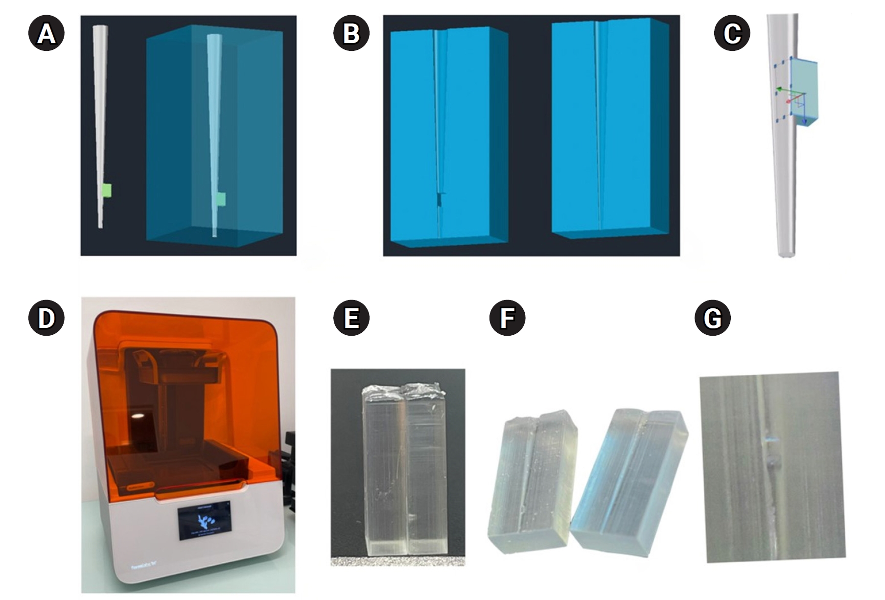
This study aimed to evaluate whether continuous irrigation with larger volumes or allowing sodium hypochlorite (NaOCl) resting time is more critical for pulp tissue dissolution using a controlled artificial root canal system.
Methods
A three-dimensional printed artificial root canal with a lateral canal in the apical third was fabricated. Standardized bovine pulp tissue specimens were inserted, and three irrigation protocols were tested: group A (continuous NaOCl irrigation at 1 mL/min via syringe pump), group B (intermittent NaOCl irrigation with 0.1 mL and a 3-minute resting period), and group C (control, saline irrigation). The time for complete dissolution and the total NaOCl volume were recorded.
Results
Complete dissolution occurred in groups A and B, with significant differences in NaOCl volume and time (p < 0.05). In group A, complete dissolution was consistently observed after the 6th irrigation cycle, corresponding to a total NaOCl volume of 6.0 ± 0.66 mL per test. The average time required for complete dissolution in this group was 6 ± 0.66 minutes. In group B, complete dissolution occurred after the 4th cycle, with a total NaOCl volume of 0.4 ± 0.06 mL per test and a mean dissolution time of 12.6 ± 1.8 minutes.
Conclusions
NaOCl volume and exposure time significantly influence pulp tissue dissolution.
- 2,084 View
- 178 Download
- Comparison of remineralization in caries-affected dentin using calcium silicate, glass ionomer cement, and resin-modified glass ionomer cement: an in vitro study
- Kwanchanok Youcharoen, Onwara Akkaratham, Papichaya Intajak, Pipop Saikaew, Sirichan Chiaraputt
- Restor Dent Endod 2025;50(4):e37. Published online November 14, 2025
- DOI: https://doi.org/10.5395/rde.2025.50.e37
-
Abstract
PDFPubReaderePub
- Objectives
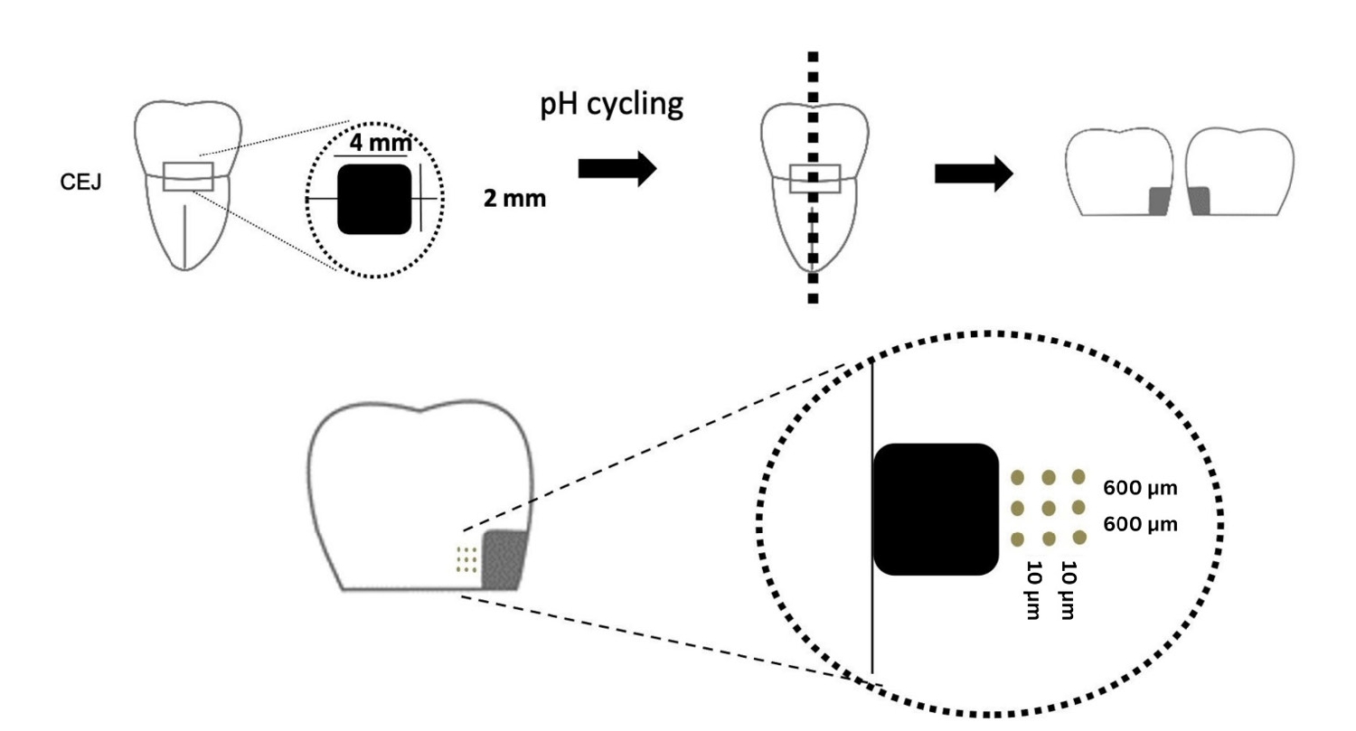
This study evaluated the ability of calcium silicate cement (CSC) as a remineralizing agent compared with conventional glass ionomer cement (GIC) and resin-modified GIC (RMGIC) to remineralize artificial caries-affected dentin.
Methods
Twenty-five class V cavities were prepared on extracted human third molars. Twenty teeth underwent artificial caries induction. The remaining five teeth with sound dentin serve as the positive control. The twenty demineralized teeth were subdivided into four groups (n = 5): carious dentin without restoration (negative control [NC]), carious dentin restored with CSC (Biodentine, Septodont), carious dentin restored with GI (Fuji IX, GC Corporation), and carious dentin restored with RMGIC (Fuji II LC, GC Corporation). Following restoration, the specimens were stored in artificial saliva for 7 days. The elastic modulus was evaluated by a nanoindentation test. The mineral composition was analyzed by scanning electron microscopy-energy-dispersive X-ray spectroscopy (SEM-EDX), and the mineral composition at the dentin-material interface.
Results
CSC had a higher modulus of elasticity compared to GI, RMGI, and NC groups (p < 0.05). Higher calcium and phosphorus content was observed under CSC restorations, as indicated by SEM-EDX examination, which may lead to better remineralization.
Conclusions
Compared to GI and RMGI, CSC showed the best remineralization and mechanical reinforcement in caries-affected dentin, indicating CSC for use in minimally invasive restorative dentistry. -
Citations
Citations to this article as recorded by- Comparison of mineral precipitation, elemental release, pH change and cytotoxicity of calcium-silicate cements and an experimental resin-modified glass ionomer cement containing bioactive glass
Wisitsin Potiprapanpong , Parichart Naruphontjirakul, Naruporn Monmaturapoj, Siriporn Tanodekaew, Somruethai Channasanon, Arnit Toneluck, Somying Patntirapong, Piyaphong Panpisut
Biomaterial Investigations in Dentistry.2026; 13: 337. CrossRef
- Comparison of mineral precipitation, elemental release, pH change and cytotoxicity of calcium-silicate cements and an experimental resin-modified glass ionomer cement containing bioactive glass
- 2,555 View
- 245 Download
- 1 Crossref
- Evaluation of platelet concentrates in regenerative endodontics: a systematic review and meta-analysis
- Anna Tsiolaki, Dimitrios Theocharis, Nikolaos Tsitsipas, Anastasia Fardi, Konstantinos Kodonas
- Restor Dent Endod 2025;50(4):e38. Published online November 28, 2025
- DOI: https://doi.org/10.5395/rde.2025.50.e38
-
Abstract
PDF
 Supplementary MaterialPubReaderePub
 - Objectives
The aim of this systematic review is to compare the effectiveness of advanced platelet concentrates as regenerative endodontic therapeutic alternatives to blood clot (BC) revascularization in immature permanent necrotic teeth.
Methods
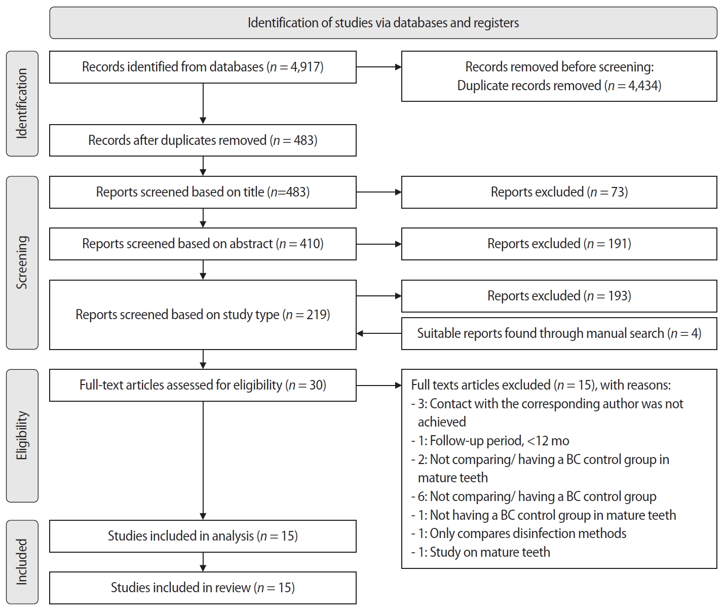
Randomized controlled trials (RCTs) comparing regenerative endodontic therapies using platelet-rich plasma (PRP), platelet-rich fibrin (PRF), or platelet pellet (PP) with the BC revascularization approach in immature permanent necrotic teeth were systematically searched in PubMed, Scopus, Cochrane Library, and Web of Science until May 2025. Data was extracted and analyzed both qualitatively and quantitatively. Study quality was assessed using the Cochrane Risk of Bias tool. A meta-analysis was conducted using IBM SPSS software (version 29.0), with success rates expressed as risk ratios and 95% confidence intervals (CIs).
Results
The initial search yielded 4,917 studies. After removing duplicates and applying eligibility criteria, 15 RCTs were included. Meta-analysis indicated no significant difference in the risk ratio (RR), as the BC method has similar success rates with PRP (10 studies; RR = 1.01; 95% CI, 0.94–1.09; p = 0.76) and PRF (8 studies; RR = 0.98; 95% CI, 0.89–1.08; p = 0.65) at 12 months. The primary outcomes evaluated were based on clinical and radiographic success.
Conclusions
Current evidence suggests PRP, PRF, and BC are all effective in treating immature permanent necrotic teeth with similar success rates. However, further research is needed to assess long-term outcomes. -
Citations
Citations to this article as recorded by- Longitudinal periapical radiographic evaluation of apexification, vital pulpotomy, and revascularization in immature permanent teeth: a retrospective comparative study
Xiaona Sun, Kailing Zhu
Frontiers in Bioengineering and Biotechnology.2026;[Epub] CrossRef
- Longitudinal periapical radiographic evaluation of apexification, vital pulpotomy, and revascularization in immature permanent teeth: a retrospective comparative study
- 2,117 View
- 109 Download
- 1 Web of Science
- 1 Crossref
- Difference in light transmittance and depth of cure of flowable composite depending on tooth thickness: an in vitro experimental study
- Seong-Pyo Bae, Myung-Jin Lee, Kyung-San Min, Mi-Kyung Yu, Kwang-Won Lee
- Restor Dent Endod 2025;50(4):e39. Published online November 28, 2025
- DOI: https://doi.org/10.5395/rde.2025.50.e39
-
Abstract
PDFPubReaderePub
- Objectives
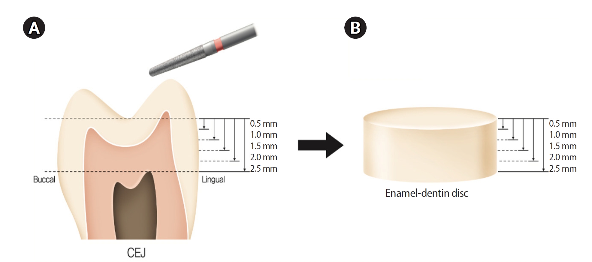
This study aimed to quantify light attenuation through varying tooth thicknesses and its impact on the depth of cure of composite resin.
Methods
Twenty extracted premolars were used to create enamel-dentin discs that were sanded progressively in 0.5 mm increments from 2.5 mm to 0.5 mm. Light irradiance was measured with and without tooth specimens to evaluate light transmittance. Resin was cured beneath different thicknesses, and the depth of cure was assessed using the Vickers hardness test.
Results
The results demonstrated that light transmittance significantly decreased as tooth thickness increased (p < 0.01), leading to reduced resin polymerization. In the 2.0-mm and 2.5-mm tooth thickness groups, the depth of cure was significantly lower than in the control group without tooth specimens (p < 0.05).
Conclusions
Ultimately, for tooth structures exceeding 2 mm, self-cure or dual-cure resin polymerization is thought to be more efficient than light polymerization.
- 1,842 View
- 141 Download
- Resolvin E1 incorporated carboxymethyl chitosan scaffold accelerates repair of dental pulp stem cells under inflammatory conditions: a laboratory investigation
- Hemalatha P Balasubramanian, Nandini Suresh, Vishnupriya Koteeswaran, Velmurugan Natanasabapathy
- Restor Dent Endod 2025;50(4):e40. Published online November 28, 2025
- DOI: https://doi.org/10.5395/rde.2025.50.e40
-
Abstract
PDFSupplementary MaterialPubReaderePub
- Objectives
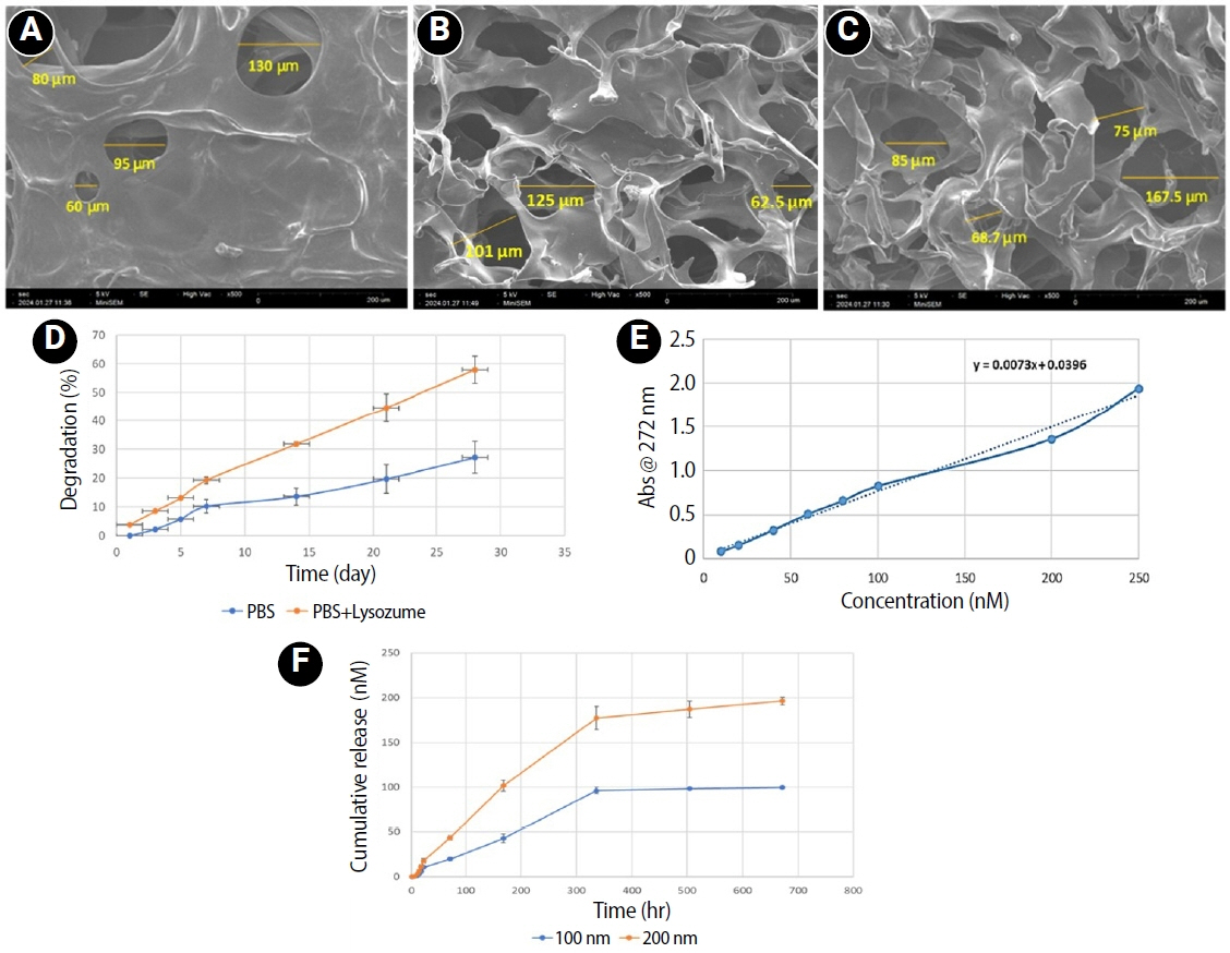
This study fabricated and characterized a resolvin E1 (RvE1)-loaded carboxymethyl chitosan (CMC) scaffold and determined its cytotoxicity and mineralization potential on inflamed human dental pulp stem cells (hDPSCs).
Methods
CMC scaffold incorporated with two concentrations of RvE1 (100 and 200 nM) was fabricated and characterized. The scaffolds’ porosity, drug release kinetics, and degradation were assessed. The impact of RvE1 on inflamed hDPSCs proliferation, proinflammatory gene expression (tumor necrosis factor alpha [TNF-α]), alkaline phosphatase activity, and alizarin red S staining was evaluated.
Results
Scanning electron microscopy analysis demonstrated a highly porous interconnected microstructure. Release kinetics showed gradual RvE1 release peaking at day 14. Cumulative degradation of the CMC scaffold at 28 days was 57.35%. Inflamed hDPSCs exposed to 200 nM RvE1-CMC scaffold exhibited significantly improved viability compared to 100 nM. Both RvE1-CMC scaffolds significantly suppressed the expression of TNF-α at 7 days. Alkaline phosphatase activity was enhanced by both RvE1 concentrations on days 7 and 14. Alizarin red staining revealed superior mineralization potential of 200 nM RvE1 on days 14 and 21.
Conclusions
This study concludes 200 nM RvE1-CMC scaffold is a promising therapy for inflamed pulp conditions, enhancing cell proliferation and biomineralization potential in inflamed hDPSCs.
- 1,256 View
- 55 Download
- Effect of moisture and pH on setting time and microhardness of three premixed calcium silicate-based root canal sealers: an in vitro experimental study
- Sooyoun Kim
- Restor Dent Endod 2025;50(4):e41. Published online November 28, 2025
- DOI: https://doi.org/10.5395/rde.2025.50.e41
-
Abstract
PDFPubReaderePub
- Objectives
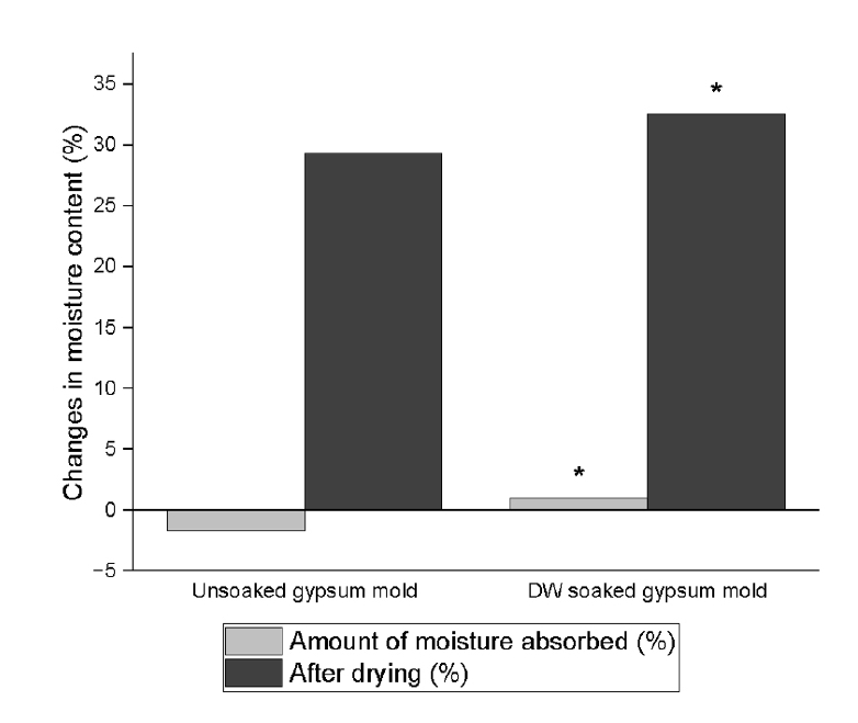
The study aimed to investigate how environmental conditions impact the setting time and microhardness of premixed calcium silicate-based sealers.
Methods
The setting time and microhardness of three sealers (Endoseal MTA [MARUCHI], One-Fil [MEDICLUS], and Well-Root ST [VERICOM]) were evaluated under four environmental conditions: unsoaked, distilled water-soaked, phosphate-buffered saline-soaked, and pH 5-soaked gypsum molds (n = 12/group/condition). The setting time was measured with Gilmore needles, and microhardness was assessed using a Vickers tester after 3 days. Welch’s analysis of variance and Games-Howell post hoc tests were used for statistical analysis.
Results
The sealer type and environmental conditions significantly influenced setting time and microhardness (p < 0.001). The initial and final setting times were the shortest in the unsoaked samples. For Endoseal MTA and One-Fil, the unsoaked condition exhibited significantly shorter setting times than the soaked conditions. Well-Root ST exhibited significantly longer setting times in acidic conditions. Surface microhardness was highest in the unsoaked group (p < 0.001). Among the soaked groups, the phosphate-buffered saline-soaked group had the lowest hardness for Endoseal MTA, whereas the pH 5-soaked group exhibited the lowest hardness for One-Fil and Well-Root ST. Endoseal MTA consistently demonstrated a lower microhardness than the other sealers (p < 0.001).
Conclusions
Moisture, pH, and solution chemistry influenced the setting time and microhardness of premixed calcium silicate sealers. Although acidic conditions generally prolong the setting time and reduce hardness, the effects vary based on the sealers used and the setting environment. -
Citations
Citations to this article as recorded by- Setting Characteristics, Solubility, Bioactivity and Interaction with Dentin of Four Calcium Silicate-Based Endodontic Sealers
Areti Dimitra Vrochari, Anastasia Agrafioti, Maria Dimitriadi, George Eliades
Journal of Functional Biomaterials.2026; 17(4): 192. CrossRef
- Setting Characteristics, Solubility, Bioactivity and Interaction with Dentin of Four Calcium Silicate-Based Endodontic Sealers
- 1,970 View
- 97 Download
- 1 Web of Science
- 1 Crossref
 First
First Prev
Prev

