-
Guided endodontics, precision and predictability: a case series of mineralized anterior teeth with follow-up cone-beam computed tomography
-
Rafael Fernández-Grisales, Wilder Javier Rojas-Gutierrez, Pamela Mejía, Carolina Berruecos-Orozco, Néstor Ríos-Osorio
-
Restor Dent Endod 2025;50(1):e4. Published online January 6, 2025
-
DOI: https://doi.org/10.5395/rde.2025.50.e4
-
-
 Abstract Abstract
 PDF PDF PubReader PubReader ePub ePub
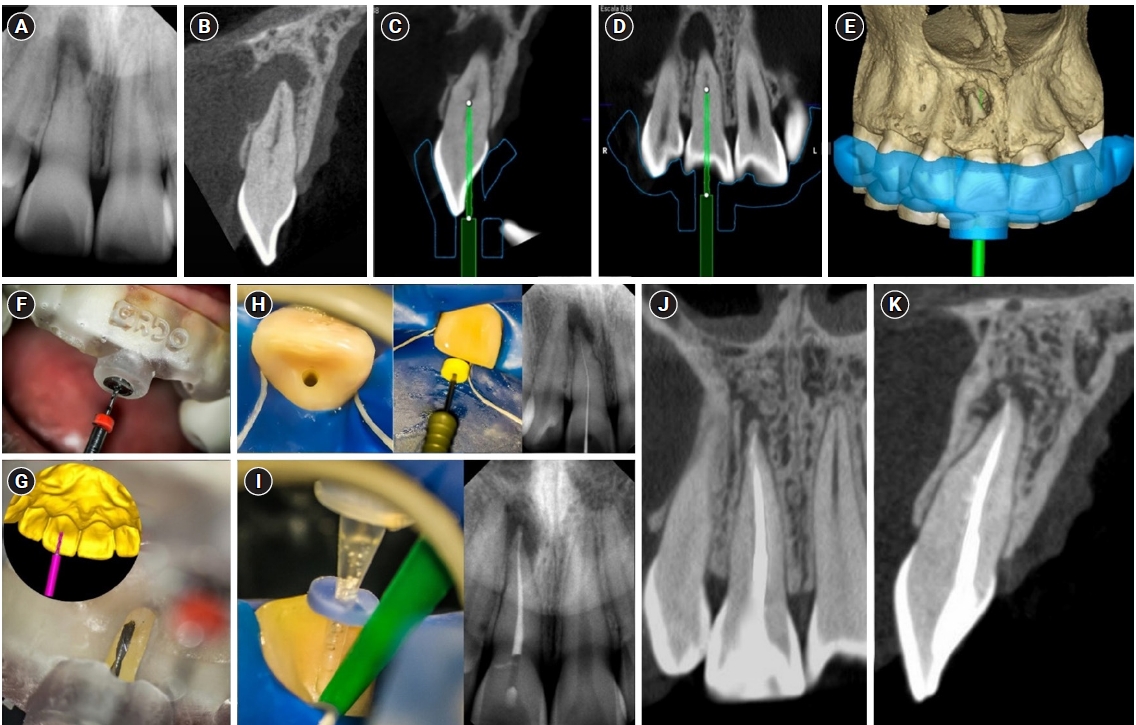
- Pulp chamber and root canal obliteration (PCO/RCO) presents a challenge for clinicians when nonsurgical endodontic treatment is indicated. Guided endodontics (GE) aims to precisely locate the root canal (RC) system while preserving as much pericervical dentin as possible. GE involves integrating cone-beam computed tomography (CBCT) of the affected tooth with a digital impression of the maxillary/mandibular arch, allowing for careful planning of the drilling path to the RC system through a three-dimensional (3D) static guide. This article reports four cases of teeth with PCO/RCO, accompanied by additional diagnoses of internal and external root resorption and horizontal tooth fracture, all successfully treated with GE. These cases highlight the clinical and radiographic success of GE treatments using CBCT, establishing this technique as a predictable approach for managing mineralized teeth.
-
Cone-beam computed tomography in endodontics: from the specific technical considerations of acquisition parameters and interpretation to advanced clinical applications
-
Néstor Ríos-Osorio, Sara Quijano-Guauque, Sandra Briñez-Rodríguez, Gustavo Velasco-Flechas, Antonieta Muñoz-Solís, Carlos Chávez, Rafael Fernandez-Grisales
-
Restor Dent Endod 2024;49(1):e1. Published online December 11, 2023
-
DOI: https://doi.org/10.5395/rde.2024.49.e1
-
-
 Abstract Abstract
 PDF PDF PubReader PubReader ePub ePub
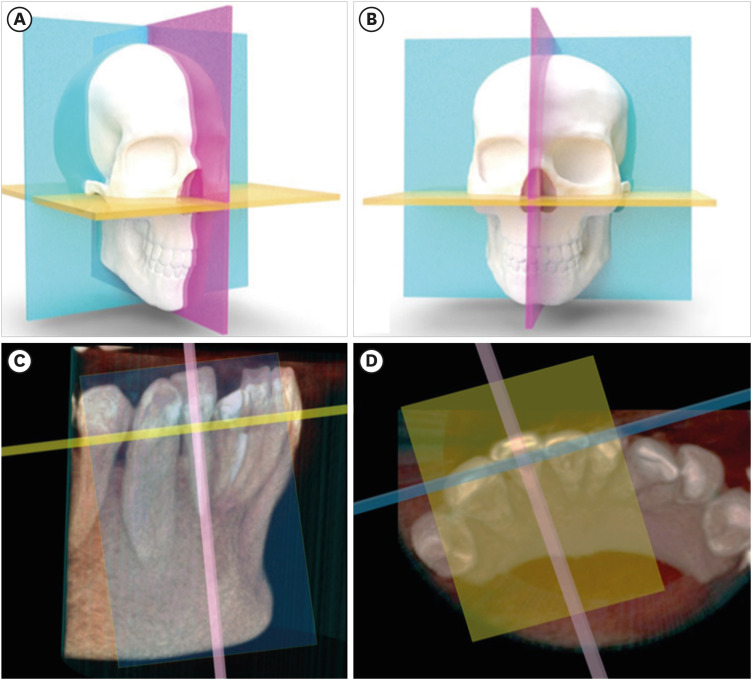
The implementation of imaging methods that enable sensitive and specific observation of anatomical structures has been a constant in the evolution of endodontic therapy. Cone-beam computed tomography (CBCT) enables 3-dimensional (3D) spatial anatomical navigation in the 3 volumetric planes (sagittal, coronal and axial) which translates into great accuracy for the identification of endodontic pathologies/conditions. CBCT interpretation consists of 2 main components: (i) the generation of specific tasks of the image and (ii) the subsequent interpretation report. A systematic and reproducible method to review CBCT scans can improve the accuracy of the interpretation process, translating into greater precision in terms of diagnosis and planning of endodontic clinical procedures. MEDLINE (PubMed), Web of Science, Google Scholar, Embase and Scopus were searched from inception to March 2023. This narrative review addresses the theoretical concepts, elements of interpretation and applications of the CBCT scan in endodontics. In addition, the contents and rationale for reporting 3D endodontic imaging are discussed. -
Citations
Citations to this article as recorded by  - Evaluation of Maxillary Sinus Pathologies in Children and Adolescents with Cleft Lip and Palate Using Cone Beam Computed Tomography: A Retrospective Study
Ayşe Çelik, Nilüfer Ersan, Senem Selvi-Kuvvetli
The Cleft Palate Craniofacial Journal.2025;[Epub] CrossRef - Machine Learning Models in the Detection of MB2 Canal Orifice in CBCT Images
Shishir Shetty, Meliz Yuvali, Ilker Ozsahin, Saad Al-Bayatti, Sangeetha Narasimhan, Mohammed Alsaegh, Hiba Al-Daghestani, Raghavendra Shetty, Renita Castelino, Leena R David, Dilber Uzun Ozsahin
International Dental Journal.2025; 75(3): 1640. CrossRef - Early diagnosis of acute lymphoblastic leukemia utilizing clinical, radiographic, and dental age indicators
Rehab F Ghouraba, Shaimaa S. EL-Desouky, Mohamed R. El-Shanshory, Ibrahim A. Kabbash, Nancy M. Metwally
Scientific Reports.2025;[Epub] CrossRef - Tomographic evaluation of apexogenesis with human treated dentin matrix in young permanent molars: a split-mouth randomized controlled clinical trial
Nora M. Abo Shanady, Nahed A. Abo Hamila, Gamal M. El Maghraby, Rehab F. Ghouraba
BMC Oral Health.2025;[Epub] CrossRef - The Integration of Cone Beam Computed Tomography, Artificial Intelligence, Augmented Reality, and Virtual Reality in Dental Diagnostics, Surgical Planning, and Education: A Narrative Review
Aida Meto, Gerta Halilaj
Applied Sciences.2025; 15(11): 6308. CrossRef - Healing Outcomes of Through‐And‐Through Bone Defects in Periapical Surgery: A Systematic Review and Meta‐Analysis
Bibi Fatima, Farhan Raza Khan, Syeda Abeerah Tanveer
Australian Endodontic Journal.2025; 51(2): 518. CrossRef - Genotoxic and cytotoxic effects of cone beam computed tomography on exfoliated epithelial cells in different age groups
Maged Bakr, Fatma Ata, Asmaa Saleh Elmahdy, Bassant Mowafey
BMC Oral Health.2025;[Epub] CrossRef - Bridging the gap in aberrant root canal systems: Case series
Seethalakshmi Tamizhselvan, Diana Davidson, Srinivasan Manali Ramakrishnan
Journal of Conservative Dentistry and Endodontics.2025; 28(8): 833. CrossRef - IMAGING TECHNIQUES IN ENDODONTIC DIAGNOSIS: A REVIEW OF LITERATURE
Mihaela Salceanu, Anca Melian , Tudor Hamburda , Cristina Antohi , Corina Concita , Claudiu Topoliceanu , Cristian Levente Giuroiu
Romanian Journal of Oral Rehabilitation.2025; 17(1): 705. CrossRef - A Three-rooted Deciduous Second Molar in a 13-year-old Caucasian Female
Daniel Traub, Robert Walsh, Colleen Ahern
International Journal of Medical Case Reports.2025; 4(3): 51. CrossRef - Bildgebung im ZMK-Bereich – aber in welcher Reihenfolge?
Rainer Lutz
Zahnmedizin up2date.2024; 18(04): 297. CrossRef - Cone-beam computed tomography evaluation of shaping ability of kedo-S square and fanta AF™ baby rotary files compared to manual K-files in root canal preparation of primary anterior teeth
Shaimaa S. El-Desouky, Bassem N. El Fahl, Ibrahim A. Kabbash, Shimaa M. Hadwa
Clinical Oral Investigations.2024;[Epub] CrossRef - Analysis of Endodontic Successes and Failures in the Removal of Fractured Endodontic Instruments during Retreatment: A Systematic Review, Meta-Analysis, and Trial Sequential Analysis
Mario Dioguardi, Corrado Dello Russo, Filippo Scarano, Fariba Esperouz, Andrea Ballini, Diego Sovereto, Mario Alovisi, Angelo Martella, Lorenzo Lo Muzio
Healthcare.2024; 12(14): 1390. CrossRef
-
12,961
View
-
560
Download
-
14
Web of Science
-
13
Crossref
|


