-
Micro-computed tomographic evaluation of single-cone obturation with three sealers
-
Sahar Zare, Ivy Shen, Qiang Zhu, Chul Ahn, Carolyn Primus, Takashi Komabayashi
-
Restor Dent Endod 2021;46(2):e25. Published online April 16, 2021
-
DOI: https://doi.org/10.5395/rde.2021.46.e25
-
-
 Abstract Abstract
 PDF PDF PubReader PubReader ePub ePub
- Objectives
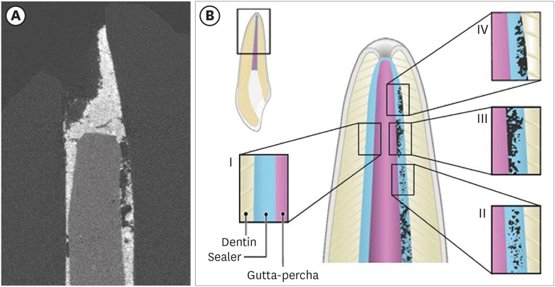
This study used micro-computed tomography (µCT) to compare voids and interfaces in single-cone obturation among AH Plus, EndoSequence BC, and prototype surface pre-reacted glass ionomer (S-PRG) sealers and to determine the percentage of sealer contact at the dentin and gutta-percha (GP) interfaces. Materials and MethodsFifteen single-rooted human teeth were shaped using ProTaper NEXT size X5 rotary files using 2.5% NaOCl irrigation. Roots were obturated with a single-cone ProTaper NEXT GP point X5 with AH Plus, EndoSequence BC, or prototype S-PRG sealer (n = 5/group). ResultsThe volumes of GP, sealer, and voids were measured in the region of 0–2, 2–4, 4–6, and 6–8 mm from the apex, using image analysis of sagittal µCT scans. GP volume percentages were: AH Plus (75.5%), EndoSequence BC (87.3%), and prototype S-PRG (94.4%). Sealer volume percentages were less: AH Plus (14.3%), EndoSequence BC (6.8%), and prototype S-PRG (4.6%). Void percentages were AH Plus (10.1%), EndoSequence BC (5.9%), and prototype S-PRG (1.0%). Dentin-sealer contact ratios of AH Plus, EndoSequence BC, and prototype S-PRG groups were 82.4% ± 6.8%, 71.6% ± 25.3%, and 70.2% ± 9.4%, respectively. GP-sealer contact ratios of AH Plus, EndoSequence BC, and prototype S-PRG groups were 65.6% ± 29.1%, 80.7% ± 25.8%, and 87.0% ± 8.6%, respectively. ConclusionsPrototype S-PRG sealer created a low-void obturation, similar to EndoSequence BC sealer with similar dentin-sealer contact (> 70%) and GP-sealer contact (> 80%). Prototype S-PRG sealer presented comparable filling quality to EndoSequence BC sealer.
-
Citations
Citations to this article as recorded by  - Assessment of gap areas of root filling techniques in teeth with 3D-printed different configurations of C-shaped root canals: a micro-computed tomography study
Tuba Gok, Adem Gok, Haydar Onur Aciksoz
BMC Oral Health.2025;[Epub] CrossRef - Assessment of isthmus filling using two obturation techniques performed by students with different levels of clinical experience
Yang Yu, Chong-Yang Yuan, Xing-Zhe Yin, Xiao-Yan Wang
Journal of Dental Sciences.2024; 19(1): 169. CrossRef - Micro-CT determination of the porosity of two tricalcium silicate sealers applied using three obturation techniques
Jinah Kim, Kali Vo, Gurmukh S. Dhaliwal, Aya Takase, Carolyn Primus, Takashi Komabayashi
Journal of Oral Science.2024; 66(3): 163. CrossRef - Ex-vivo evaluation of clinically-set hydraulic sealers used with different canal dryness protocols and obturation techniques: a randomized clinical trial
Nawar Naguib Nawar, Mohamed Mohamed Elashiry, Ahmed El Banna, Shehabeldin Mohamed Saber, Edgar Schäfer
Clinical Oral Investigations.2024;[Epub] CrossRef - Hydraulic (Single Cone) Versus Thermogenic (Warm Vertical Compaction) Obturation Techniques: A Systematic Review
Haytham S Jaha
Cureus.2024;[Epub] CrossRef - Sealing ability of various endodontic sealers with or without ethylenediaminetetraacetic acid (EDTA) treatment on bovine root canal
Yusuke AIGAMI, Tomofumi SAWADA, Shunsuke SHIMIZU, Akiko ASANO, Mamoru NODA, Shinji TAKEMOTO
Dental Materials Journal.2024; 43(3): 420. CrossRef - A Literature Review of the Effect of Heat on the Physical-Chemical Properties of Calcium Silicate–Based Sealers
Israa Ashkar, José Luis Sanz, Leopoldo Forner, James Ghilotti, María Melo
Journal of Endodontics.2024; 50(8): 1044. CrossRef - Assessment of the Prevalence of Head Lice Infestation and Parents’ Attitudes Towards Its Management: A School-based Epidemiological Study in İstanbul, Türkiye
Özben Özden, İnci Timur, Hale Ezgi Açma, Duygu Şimşekli, Barış Gülerman, Özgür Kurt
Turkish Journal of Parasitology.2023; 47(2): 112. CrossRef - Calcium-doped zinc oxide nanocrystals as an innovative intracanal medicament: a pilot study
Gabriela Leite de Souza, Thamara Eduarda Alves Magalhães, Gabrielle Alves Nunes Freitas, Nelly Xiomara Alvarado Lemus, Gabriella Lopes de Rezende Barbosa, Anielle Christine Almeida Silva, Camilla Christian Gomes Moura
Restorative Dentistry & Endodontics.2022;[Epub] CrossRef - Micro‐CT assessment of gap‐containing areas along the gutta‐percha‐sealer interface in oval‐shaped canals
Gustavo De‐Deus, Gustavo O. Santos, Iara Zamboni Monteiro, Daniele M. Cavalcante, Marco Simões‐Carvalho, Felipe G. Belladonna, Emmanuel J. N. L. Silva, Erick M. Souza, Raphael Licha, Carla Zogheib, Marco A. Versiani
International Endodontic Journal.2022; 55(7): 795. CrossRef - A critical analysis of research methods and experimental models to study root canal fillings
Gustavo De‐Deus, Erick Miranda Souza, Emmanuel João Nogueira Leal Silva, Felipe Gonçalves Belladonna, Marco Simões‐Carvalho, Daniele Moreira Cavalcante, Marco Aurélio Versiani
International Endodontic Journal.2022; 55(S2): 384. CrossRef - Use of micro-CT to examine effects of heat on coronal obturation
Ivy Shen, Joan Daniel, Kali Vo, Chul Ahn, Carolyn Primus, Takashi Komabayashi
Journal of Oral Science.2022; 64(3): 224. CrossRef - Obturation of Root Canals By Vertical Condensation of Gutta-Percha – Benefits and Pitfalls
Calkovsky Bruno, Slobodnikova Ladislava, Bacinsky Martin, Janickova Maria
Acta Medica Martiniana.2021; 21(3): 103. CrossRef
-
261
View
-
11
Download
-
11
Web of Science
-
13
Crossref
-
Antifungal effects of synthetic human β-defensin 3-C15 peptide
-
Sang-Min Lim, Ki-Bum Ahn, Christine Kim, Jong-Won Kum, Hiran Perinpanayagam, Yu Gu, Yeon-Jee Yoo, Seok Woo Chang, Seung Hyun Han, Won-Jun Shon, Woocheol Lee, Seung-Ho Baek, Qiang Zhu, Kee-Yeon Kum
-
Restor Dent Endod 2016;41(2):91-97. Published online March 17, 2016
-
DOI: https://doi.org/10.5395/rde.2016.41.2.91
-
-
 Abstract Abstract
 PDF PDF PubReader PubReader ePub ePub
- Objectives
The purpose of this ex vivo study was to compare the antifungal activity of a synthetic peptide consisting of 15 amino acids at the C-terminus of human β-defensin 3 (HBD3-C15) with calcium hydroxide (CH) and Nystatin (Nys) against Candida albicans (C. albicans) biofilm. Materials and MethodsC. albicans were grown on cover glass bottom dishes or human dentin disks for 48 hr, and then treated with HBD3-C15 (0, 12.5, 25, 50, 100, 150, 200, and 300 µg/mL), CH (100 µg/mL), and Nys (20 µg/mL) for 7 days at 37℃. On cover glass, live and dead cells in the biomass were measured by the FilmTracer Biofilm viability assay, and observed by confocal laser scanning microscopy (CLSM). On dentin, normal, diminished and ruptured cells were observed by field-emission scanning electron microscopy (FE-SEM). The results were subjected to a two-tailed t-test, a one way analysis variance and a post hoc test at a significance level of p = 0.05. ResultsC. albicans survival on dentin was inhibited by HBD3-C15 in a dose-dependent manner. There were fewer aggregations of C. albicans in the groups of Nys and HBD3-C15 (≥ 100 µg/mL). CLSM showed C. albicans survival was reduced by HBD3-C15 in a dose dependent manner. Nys and HBD3-C15 (≥ 100 µg/mL) showed significant fungicidal activity compared to CH group (p < 0.05). ConclusionsSynthetic HBD3-C15 peptide (≥ 100 µg/mL) and Nys exhibited significantly higher antifungal activity than CH against C. albicans by inhibiting cell survival and biofilm.
-
Citations
Citations to this article as recorded by  - Current status of antimicrobial peptides databases and computational tools for optimization
Madhulika Jha, Akash Nautiyal, Kumud Pant, Navin Kumar
Environment Conservation Journal.2025; 26(1): 281. CrossRef - Anti-fungal peptides: an emerging category with enthralling therapeutic prospects in the treatment of candidiasis
Jyoti Sankar Prusty, Ashwini Kumar, Awanish Kumar
Critical Reviews in Microbiology.2024; : 1. CrossRef - Harnessing antimicrobial peptides in endodontics
Xinzi Kong, Vijetha Vishwanath, Prasanna Neelakantan, Zhou Ye
International Endodontic Journal.2024; 57(7): 815. CrossRef - Human β-defensins and their synthetic analogs: Natural defenders and prospective new drugs of oral health
Mumian Chen, Zihe Hu, Jue Shi, Zhijian Xie
Life Sciences.2024; 346: 122591. CrossRef - Candida albicans Virulence Factors and Pathogenicity for Endodontic Infections
Yeon-Jee Yoo, A Reum Kim, Hiran Perinpanayagam, Seung Hyun Han, Kee-Yeon Kum
Microorganisms.2020; 8(9): 1300. CrossRef - Innate Inspiration: Antifungal Peptides and Other Immunotherapeutics From the Host Immune Response
Derry K. Mercer, Deborah A. O'Neil
Frontiers in Immunology.2020;[Epub] CrossRef - Human salivary proteins and their peptidomimetics: Values of function, early diagnosis, and therapeutic potential in combating dental caries
Kun Wang, Xuedong Zhou, Wei Li, Linglin Zhang
Archives of Oral Biology.2019; 99: 31. CrossRef - Endodontic biofilms: contemporary and future treatment options
Yeon-Jee Yoo, Hiran Perinpanayagam, Soram Oh, A-Reum Kim, Seung-Hyun Han, Kee-Yeon Kum
Restorative Dentistry & Endodontics.2019;[Epub] CrossRef - Bioactive Peptides Against Fungal Biofilms
Karen G. N. Oshiro, Gisele Rodrigues, Bruna Estéfani D. Monges, Marlon Henrique Cardoso, Octávio Luiz Franco
Frontiers in Microbiology.2019;[Epub] CrossRef - Anticandidal Potential of Stem Bark Extract from Schima superba and the Identification of Its Major Anticandidal Compound
Chun Wu, Hong-Tan Wu, Qing Wang, Guey-Horng Wang, Xue Yi, Yu-Pei Chen, Guang-Xiong Zhou
Molecules.2019; 24(8): 1587. CrossRef - Synthetic Human β Defensin-3-C15 Peptide in Endodontics: Potential Therapeutic Agent in Streptococcus gordonii Lipoprotein-Stimulated Human Dental Pulp-Derived Cells
Yeon-Jee Yoo, Hiran Perinpanayagam, Jue-Yeon Lee, Soram Oh, Yu Gu, A-Reum Kim, Seok-Woo Chang, Seung-Ho Baek, Kee-Yeon Kum
International Journal of Molecular Sciences.2019; 21(1): 71. CrossRef - Candida Infections and Therapeutic Strategies: Mechanisms of Action for Traditional and Alternative Agents
Giselle C. de Oliveira Santos, Cleydlenne C. Vasconcelos, Alberto J. O. Lopes, Maria do S. de Sousa Cartágenes, Allan K. D. B. Filho, Flávia R. F. do Nascimento, Ricardo M. Ramos, Emygdia R. R. B. Pires, Marcelo S. de Andrade, Flaviane M. G. Rocha, Cristi
Frontiers in Microbiology.2018;[Epub] CrossRef - Perspectives for clinical use of engineered human host defense antimicrobial peptides
María Eugenia Pachón-Ibáñez, Younes Smani, Jerónimo Pachón, Javier Sánchez-Céspedes
FEMS Microbiology Reviews.2017; 41(3): 323. CrossRef - The synthetic human beta-defensin-3 C15 peptide exhibits antimicrobial activity against Streptococcus mutans, both alone and in combination with dental disinfectants
Ki Bum Ahn, A. Reum Kim, Kee-Yeon Kum, Cheol-Heui Yun, Seung Hyun Han
Journal of Microbiology.2017; 55(10): 830. CrossRef - Antibiofilm peptides against oral biofilms
Zhejun Wang, Ya Shen, Markus Haapasalo
Journal of Oral Microbiology.2017; 9(1): 1327308. CrossRef - Humanβ-Defensin 3 Reduces TNF-α-Induced Inflammation and Monocyte Adhesion in Human Umbilical Vein Endothelial Cells
Tianying Bian, Houxuan Li, Qian Zhou, Can Ni, Yangheng Zhang, Fuhua Yan
Mediators of Inflammation.2017; 2017: 1. CrossRef - Antifungal Effects of Synthetic Human Beta-defensin-3-C15 Peptide on Candida albicans –infected Root Dentin
Yeon-Jee Yoo, Ikyung Kwon, So-Ram Oh, Hiran Perinpanayagam, Sang-Min Lim, Ki-Bum Ahn, Yoon Lee, Seung-Hyun Han, Seok-Woo Chang, Seung-Ho Baek, Qiang Zhu, Kee-Yeon Kum
Journal of Endodontics.2017; 43(11): 1857. CrossRef - A 15-amino acid C-terminal peptide of beta-defensin-3 inhibits bone resorption by inhibiting the osteoclast differentiation and disrupting podosome belt formation
Ok-Jin Park, Jiseon Kim, Ki Bum Ahn, Jue Yeon Lee, Yoon-Jeong Park, Kee-Yeon Kum, Cheol-Heui Yun, Seung Hyun Han
Journal of Molecular Medicine.2017; 95(12): 1315. CrossRef
-
247
View
-
2
Download
-
18
Crossref
|


