-
A 48-month clinical performance of hybrid ceramic fragment restorations manufactured in CAD/CAM in non-carious cervical lesions: case report
-
Michael Willian Favoreto, Gabriel David Cochinski, Eveline Claudia Martini, Thalita de Paris Matos, Matheus Coelho Bandeca, Alessandro Dourado Loguercio
-
Restor Dent Endod 2024;49(3):e32. Published online August 5, 2024
-
DOI: https://doi.org/10.5395/rde.2024.49.e32
-
-
 Abstract Abstract
 PDF PDF PubReader PubReader ePub ePub
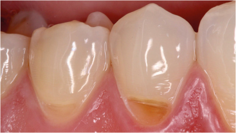
From the restorative perspective, various methods are available to prevent the progression of non-carious cervical lesions. Direct, semi-direct, and indirect composite resin techniques and indirect ceramic restorations are commonly recommended. In this context, semi-direct and indirect restoration approaches are increasingly favored, particularly as digital dentistry becomes more prevalent. To illustrate this, we present a case report demonstrating the efficacy of hybrid ceramic fragments fabricated using computer-aided design (CAD)/computer-aided manufacturing (CAM) technology and cemented with resin cement in treating non-carious cervical lesions over a 48-month follow-up period. A 24-year-old male patient sought treatment for aesthetic concerns and dentin hypersensitivity in the cervical region of the lower premolar teeth. Clinical examination confirmed the presence of two non-carious cervical lesions in the buccal region of teeth #44 and #45. The treatment plan involved indirect restoration using CAD/CAM-fabricated hybrid ceramic fragments as a restorative material. After 48 months, the hybrid ceramic material exhibited excellent adaptation and durability provided by the CAD/CAM system. This case underscores the effectiveness of hybrid ceramic fragments in restoring non-carious cervical lesions, highlighting their long-term stability and clinical success.
-
Evaluation of at-home bleaching protocol with application on different surfaces: bleaching efficacy and hydrogen peroxide permeability
-
Heloisa Forville, Michael Willian Favoreto, Michel Wendlinger, Roberta Micheten Dias, Christiane Philippini Ferreira Borges, Alessandra Reis, Alessandro D. Loguercio
-
Restor Dent Endod 2023;48(4):e33. Published online October 6, 2023
-
DOI: https://doi.org/10.5395/rde.2023.48.e33
-
-
 Abstract Abstract
 PDF PDF PubReader PubReader ePub ePub
- Objectives
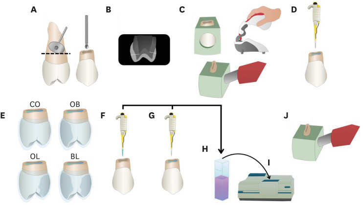
This study aimed to evaluate the bleaching efficacy and hydrogen peroxide permeability in the pulp chamber by the at-home bleaching gel in protocols applied on different dental surfaces. Materials and MethodsForty premolars were randomly into 4 groups: control group no bleaching, only application on the buccal surface (OB), only application on the lingual surface (OL) and application in buccal and lingual surfaces, simultaneously (BL). At-home bleaching gel (White Class 7.5%) was used for the procedure. The bleaching efficacy was evaluated with a digital spectrophotometer (color change in CIELAB [ΔE
ab] and CIEDE 2000 [ΔE
00] systems and Whitening Index for Dentistry [ΔWID]). The hydrogen peroxide permeability in the pulp chamber (µg/mL) was assessed using UV-Vis spectrophotometry and data were analyzed for a 1-way analysis of variance and Tukey’s test (α = 0.05). ResultsAll groups submitted to bleaching procedure showed bleaching efficacy when measured with ΔE
ab and ΔE
00 (p > 0.05). Therefore, when analyzed by ΔWID, a higher bleaching efficacy were observed for the application on the groups OB and BL (p = 0.00003). Similar hydrogen peroxide permeability was found in the pulp chambers of the teeth undergoing different protocols (p > 0.05). ConclusionsThe application of bleaching gel exclusively on the OB is sufficient to achieve bleaching efficacy, when compared to BL. Although the OL protocol demonstrated lower bleaching efficacy based on the ΔWID values, it may still be of interest and relevant in certain clinical scenarios based on individual needs, requiring clinical trials to better understand its specificities.
-
Citations
Citations to this article as recorded by  - Effect of whitening pens on hydrogen peroxide permeability in the pulp chamber, color change and surface morphology
Laryssa Mylenna Madruga Barbosa, Gabrielle Gomes Centenaro, Deisy Cristina Ferreira Cordeiro, Maria Alice de Matos Rodrigues, Letícia Condolo, Michael Willian Favoreto, Alessandra Reis, Alessandro D. Loguercio
Journal of Dentistry.2025; 154: 105595. CrossRef - Efficacy of a buccal and lingual at‐home bleaching protocol—A randomized, split‐mouth, single‐blind controlled trial
Heloisa Forville, Laís Giacomini Bernardi, Michael Willian Favoreto, Felipe Coppla, Taynara de Souza Carneiro, Fabiana Madalozzo Coppla, Alessandro D. Loguercio, Alessandra Reis
Journal of Esthetic and Restorative Dentistry.2024; 36(9): 1301. CrossRef - REANATOMIZAÇÃO DE DENTE CONOIDE ASSOCIADA A ESTÉTICA VERMELHA: RELATO DE CASO
Ana Karolayne Sousa de Morais, Daniele Fernanda Sousa Barros, Daniel Messias Limeira, Rhana Leticia de Oliveira Faria, Roberta Furtado Carvalho, Sandna Nolêto de Araújo, Laura Barbosa Santos Di Milhomem
Revista Contemporânea.2024; 4(10): e6299. CrossRef - Effect of the reduction in the exposure time to at-home bleaching gel on color change and tooth sensitivity: A systematic review and meta-analysis
Priscila Borges Gobbo de Melo, Letícia Vasconcelos Silva Souza, Lucianne Cople Maia, Guido Artemio Marañón-Vásquez, Matheus Kury, Vanessa Cavalli
Clinical Oral Investigations.2024;[Epub] CrossRef
-
372
View
-
18
Download
-
3
Web of Science
-
4
Crossref
-
Effect of medium or high concentrations of in-office dental bleaching gel on the human pulp response in the mandibular incisors
-
Douglas Augusto Roderjan, Rodrigo Stanislawczuk, Diana Gabriela Soares, Carlos Alberto de Souza Costa, Michael Willian Favoreto, Alessandra Reis, Alessandro D. Loguercio
-
Restor Dent Endod 2023;48(2):e12. Published online March 8, 2023
-
DOI: https://doi.org/10.5395/rde.2023.48.e12
-
-
 Abstract Abstract
 PDF PDF PubReader PubReader ePub ePub
- Objectives
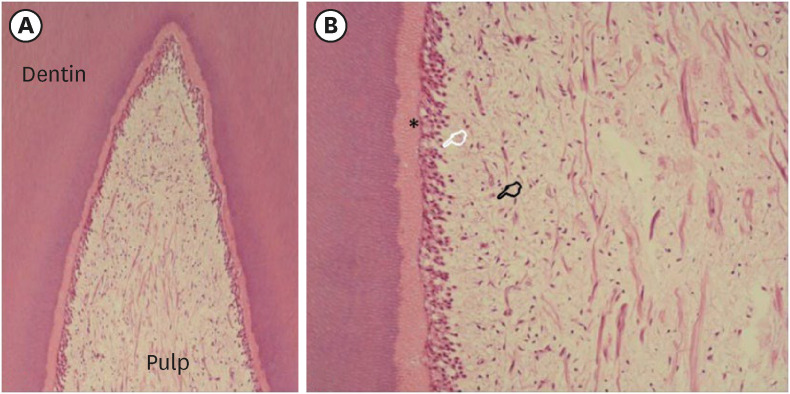
The present study evaluated the pulp response of human mandibular incisors subjected to in-office dental bleaching using gels with medium or high concentrations of hydrogen peroxide (HP). Materials and MethodsThe following groups were compared: 35% HP (HP35; n = 5) or 20% HP (HP20; n = 4). In the control group (CONT; n = 2), no dental bleaching was performed. The color change (CC) was registered at baseline and after 2 days using the Vita Classical shade guide. Tooth sensitivity (TS) was also recorded for 2 days post-bleaching. The teeth were extracted 2 days after the clinical procedure and subjected to histological analysis. The CC and overall scores for histological evaluation were evaluated by the Kruskal-Wallis and Mann-Whitney tests. The percentage of patients with TS was evaluated by the Fisher exact test (α = 0.05). ResultsThe CC and TS of the HP35 group were significantly higher than those of the CONT group (p < 0.05) and the HP20 group showed an intermediate response, without significant differences from either the HP35 or CONT group (p > 0.05). In both experimental groups, the coronal pulp tissue exhibited partial necrosis associated with tertiary dentin deposition. Overall, the subjacent pulp tissue exhibited a mild inflammatory response. ConclusionsIn-office bleaching therapies using bleaching gels with 20% or 35% HP caused similar pulp damage to the mandibular incisors, characterized by partial necrosis, tertiary dentin deposition, and mild inflammation.
-
Citations
Citations to this article as recorded by  - Can pigments of different natures interfere with the cytotoxicity from in-office bleaching?
Rafael Antonio de Oliveira Ribeiro, Beatriz Voss Martins, Marlon Ferreira Dias, Victória Peruchi, Caroline Anselmi, Igor Paulino Mendes Soares, Josimeri Hebling, Vanessa Cavalli, Carlos Alberto de Souza Costa
Odontology.2025;[Epub] CrossRef - Combined catalytic strategies applied to in-office tooth bleaching: whitening efficacy, cytotoxicity, and gene expression of human dental pulp cells in a 3D culture model
Rafael Antonio de Oliveira Ribeiro, Victória Peruchi, Igor Paulino Mendes Sores, Filipe Koon Wu Mon, Diana Gabriela Soares, Josimeri Hebling, Carlos Alberto de Souza Costa
Clinical Oral Investigations.2024;[Epub] CrossRef - Low and high hydrogen peroxide concentrations of in-office dental bleaching associated with violet light: an in vitro study
Isabela Souza Vardasca, Michael Willian Favoreto, Mylena de Araujo Regis, Taynara de Souza Carneiro, Emanuel Adriano Hul, Christiane Philippini Ferreira Borges, Alessandra Reis, Alessandro D. Loguercio, Carlos Francci
Clinical Oral Investigations.2024;[Epub] CrossRef - Evaluation of hydrogen peroxide permeability, color change, and physical–chemical properties on the in‐office dental bleaching with different mixing tip
Michael Willian Favoreto, Sibelli Olivieri Parreiras, Michel Wendlinger, Taynara De Souza Carneiro, Mariah Ignez Lenhani, Christiane Phillipini Ferreira Borges, Alessandra Reis, Alessandro D. Loguercio
Journal of Esthetic and Restorative Dentistry.2024; 36(3): 460. CrossRef - Catalysis-based approaches with biopolymers and violet LED to improve in-office dental bleaching
Rafael Antonio de Oliveira Ribeiro, Beatriz Voss Martins, Marlon Ferreira Dias, Victória Peruchi, Igor Paulino Mendes Soares, Caroline Anselmi, Josimeri Hebling, Carlos Alberto de Souza Costa
Lasers in Medical Science.2024;[Epub] CrossRef - Feasibility and Safety of Adopting a New Approach in Delivering a 450 nm Blue Laser with a Flattop Beam Profile in Vital Tooth Whitening. A Clinical Case Series with an 8-Month Follow-Up
Reem Hanna, Ioana Cristina Miron, Stefano Benedicenti
Journal of Clinical Medicine.2024; 13(2): 491. CrossRef - Hydrogen Peroxide in the Pulp Chamber and Color Change in Maxillary Anterior Teeth After In-Office Bleaching
Alexandra Mena-Serrano, Sandra Sanchez, María G. Granda-Albuja, Michael Willian Favoreto, Taynara de Souza Carneiro, Deisy Cristina Ferreira Cordeiro, Alessandro D. Loguercio, Alessandra Reis
Brazilian Dental Journal.2024;[Epub] CrossRef - Influence of coating dental enamel with a TiF4-loaded polymeric primer on the adverse effects caused by a bleaching gel with 35% H2O2
Victória Peruchi, Rafael Antonio de Oliveira Ribeiro, Igor Paulino Mendes Soares, Lídia de Oliveira Fernandes, Juliana Rios de Oliveira, Maria Luiza Barucci Araújo Pires, Josimeri Hebling, Diana Gabriela Soares, Carlos Alberto de Souza Costa
Journal of the Mechanical Behavior of Biomedical Materials.2024; 153: 106497. CrossRef
-
346
View
-
24
Download
-
7
Web of Science
-
8
Crossref
|


