-
Reference values for pulp oxygen saturation as a diagnostic tool in endodontics: a systematic review and meta-analysis
-
Paula Lambert, Sergio Augusto Quevedo Miguens, Caroline Solda, Juliana Tomaz Sganzerla, Leandro Azambuja Reichert, Carlos Estrela, Fernando Branco Barletta
-
Restor Dent Endod 2020;45(4):e48. Published online October 5, 2020
-
DOI: https://doi.org/10.5395/rde.2020.45.e48
-
-
 Abstract Abstract
 PDF PDF PubReader PubReader ePub ePub
- Objectives
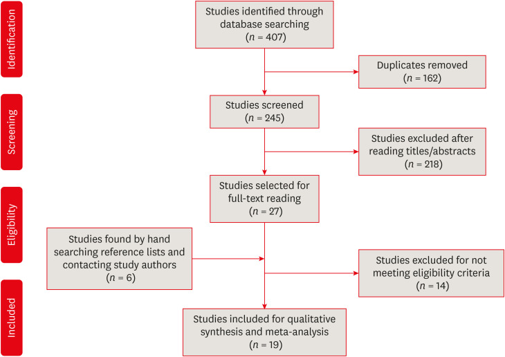
This systematic review aimed to identify mean oxygen saturation values (SpO2) using pulse oximetry in permanent maxillary anterior teeth. Materials and MethodsThe MEDLINE, Scientific Electronic Library Online, Cochrane Central Register of Controlled Trials, EMBASE, and Literatura Latino Americana em Ciências da Saúde electronic databases were searched. Combinations and variations of “oximetry” AND “dental pulp test” were used as search terms. Studies reporting means and standard deviations of SpO2 values were included. Two reviewers independently extracted data following the Preferred Reporting Items for Systematic Reviews and Meta-Analyses checklist. Heterogeneity was assessed using the I2 statistic, and all analyses were performed using R software. Study quality was assessed using the Quality Assessment of Diagnostic Accuracy Studies-2 tool and the Newcastle-Ottawa scale. ResultsOf the 251 studies identified, 19 met the eligibility criteria and were included (total sample, 4,541 teeth). In the meta-analysis, the mean SpO2 values were 84.94% (95% confidence interval [CI], 84.85%–85.04%) for the central incisors, 89.29% (95% CI, 89.22%–89.35%) for the lateral incisors, and 89.20% (95% CI, 89.05%–89.34%) for the canines. The studies were predominantly low-quality due to the high risk of bias associated with the index test, unclear risk regarding patient selection, and concerns about outcome assessment. ConclusionsAlthough most studies were low-quality, the oxygen saturation levels in normal pulp could be established (minimum saturation, 77.52%). Despite the risk of bias of the included studies, the reference values reported herein are clinically relevant for assessments of changes in pulp status. Trial RegistrationInternational Prospective Register of Systematic Reviews Identifier: CRD42018085598
-
Citations
Citations to this article as recorded by  - Lower dental pulp oxygen saturation in children with molar incisor hypomineralization: a cross-sectional study
Jade de Souza CAVALCANTE, Carlos ESTRELA, Fabrício Kitazono de CARVALHO, Francisco Wanderley Garcia de PAULA-SILVA, Manoel Damião de SOUSA-NETO, Kelly Fernanda MOLENA, Alexandra Mussolino de QUEIROZ
Brazilian Oral Research.2026;[Epub] CrossRef - Reference Values for Pulse Oximetry Testing in Permanent Teeth: A Systematic Review and Meta‐Analysis
Lilian Tietz, Theodoro Weissheimer, Cassiano Kuchenbecker Rösing, Marcus Vinicius Reis Só
International Endodontic Journal.2026;[Epub] CrossRef - Clinical Validation of Smartphone-Enabled Pulse Oximetry for Objective Pulp Vitality Assessment: A Diagnostic Accuracy Study
Celso Luiz Caldeira, Stephanie Isabel Diaz Zamalloa, Claudia Regina Guimaro Sakitani, Fernando Branco Barletta, Marinella Holzhausen
Journal of Endodontics.2025; 51(12): 1752. CrossRef - Laser Doppler Flowmetry and Continuous Tissue Oxygenation Monitoring: Best of Vitality Tests?
Herman J. J. Roeykens, Rani D’haese, Wolfgang Jacquet, Roeland J. G. De Moor, Stefan Vandeweghe
Oral.2025; 5(4): 83. CrossRef - Diagnostic accuracy of Transmitted-light plethysmography for the assessment of pulpal circulation in traumatized young permanent incisors
Satoko Kakino, Hiroaki Ohki, Kaori Kohi, Yuko Matsumura, Tsutomu Iwamoto
Scientific Reports.2025;[Epub] CrossRef - Future trends in endodontics
Foo Suanhow, Tawil Bill
Journal of Applied Biotechnology & Bioengineering.2024; 11(1): 1. CrossRef - Assessment of Pulpal Oxygen Saturation in Caries-free and Carious Maxillary Primary Central Incisors Using a Customized Dental Pulse Oximeter
Kranthi Reddy Kanumuru, Nancy Solomon, Hemalatha Ramkumar, Shankar Paulindraraj, Trophimus Gnanabagyan Jayakaran, Senthil Dakshinamoorthy
International Journal of Clinical Pediatric Dentistry.2023; 16(4): 560. CrossRef - Age-Related Variation of Pulpal Oxygen Saturation in Healthy Primary and Permanent Teeth in Children: A Clinical Study
Andreea Igna, Darian Rusu, Emilia Ogodescu, Ștefania Dinu, Marius Boariu, Adrian Voicu, Ștefan-Ioan Stratul
Journal of Clinical Medicine.2022; 12(1): 170. CrossRef - Pulp oxygen saturation measurement as a diagnostic tool for assessing pulp status in primary teeth: A systematic review and meta-analysis
Kanamarlapudi Venkata Saikiran, Deepa Gurunathan, Sainath Reddy Elicherla, Sreekanth Kumar Mallineni, Sivakumar Nuvvula
Journal of Indian Society of Pedodontics and Preventive Dentistry.2022; 40(4): 349. CrossRef - Diagnostic Value of Serum Chitinase‐3‐Like Protein 1 for Liver Fibrosis: A Meta‐analysis
Xiaoting Huang, Jialing Zhuang, Yongqiang Yang, Jiaxin Jian, Wen Ai, Chunyong Liu, Wenzhi Tang, Changyu Jiang, Yongshen He, Lesheng Huang, Se Peng, Jin Shui Pan
BioMed Research International.2022;[Epub] CrossRef - Assessment of Pulpal Status in Primary Teeth Following Direct Pulp Capping in an Experimental Canine Model
Andreea Igna, Cornel Igna, Mariana Ioana Miron, Larisa Schuszler, Roxana Dascălu, Mihaela Moldovan, Adrian Aristide Voicu, Carmen Darinca Todea, Marius Boariu, Maria-Alexandra Mârțu, Ștefan-Ioan Stratul
Diagnostics.2022; 12(8): 2022. CrossRef
-
3,217
View
-
43
Download
-
11
Crossref
-
Cytocompatibility and cell proliferation evaluation of calcium phosphate-based root canal sealers
-
Letícia Boldrin Mestieri, Ivana Maria Zaccara, Lucas Siqueira Pinheiro, Fernando Branco Barletta, Patrícia Maria Polli Kopper, Fabiana Soares Grecca
-
Restor Dent Endod 2020;45(1):e2. Published online November 15, 2019
-
DOI: https://doi.org/10.5395/rde.2020.45.e2
-
-
Abstract
PDFPubReaderePub
- Objectives
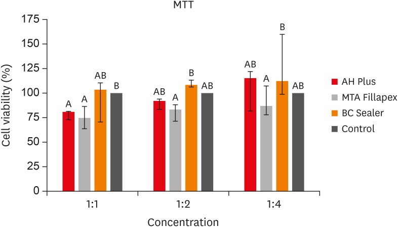
This study aimed to evaluate the cell viability and migration of Endosequence Bioceramic Root Canal Sealer (BC Sealer) compared to MTA Fillapex and AH Plus. Materials and MethodsBC Sealer, MTA Fillapex, and AH Plus were placed in contact with culture medium to obtain sealers extracts in dilution 1:1, 1:2 and 1:4. 3T3 cells were plated and exposed to the extracts. Cell viability and migration were assessed by 3-(4,5-dimethyl-thiazoyl)-2,5-diphenyl-tetrazolium bromide (MTT) and Scratch assay, respectively. Data were analyzed by Kruskal-Wallis and Dunn's test (p < 0.05). ResultsThe MTT assay revealed greater cytotoxicity for AH Plus and MTA Fillapex at 1:1 dilution when compared to control (p < 0.05). At 1:2 and 1:4 dilutions, all sealers were similar to control (p > 0.05) and MTA Fillapex was more cytotoxic than BC Sealer (p < 0.05). Scratch assay demonstrated the continuous closure of the wound according to time. At 30 hours, the control group presented closure of the wound (p < 0.05). At 36 hours, only BC Sealer presented the closure when compared to AH Plus and MTA Fillapex (p < 0.05). At 42 hours, AH Plus and MTA Fillapex showed a wound healing (p > 0.05). ConclusionsAll tested sealers demonstrated cell viability highlighting BC Sealer, which showed increased cell migration capacity suggesting that this sealer may achieve better tissue repair when compared to other tested sealers.
-
Citations
Citations to this article as recorded by - Oxidative Stress, Pro-Inflammatory Response, Cytotoxicity and Apoptosis Induced by Contemporary Endodontic Sealers in Human Periodontal Ligament Fibroblasts
Stanisław Krokosz, Virginia Ewa Lis, Sara Zięba, Mateusz Maciejczyk, Ewa Zalewska, Maria Obrycka, Edyta Gołaś, Małgorzata Żendzian-Piotrowska, Jerzy Ładny, Anna Skutnik-Radziszewska, Karol Dąbrowski, Julia Kuźmiuk, Anna Zalewska
Journal of Functional Biomaterials.2026; 17(2): 105. CrossRef - Effect of Nano-Silica on Mechanical Properties and Cytotoxicity of Calcium-Silicate-Based Root Canal Filling Materials
Hao He, Bolang Hao, Xiang Xiong, Yi Cheng, Jia Lou, Zheyu He, Dongyang Li, Zhihuan Wang, Jian Qin
Crystals.2025; 15(1): 55. CrossRef - Premixed calcium silicate-based root canal sealers have better biological properties than AH Plus: A systematic review and meta-analysis of in vivo animal studies and in vitro laboratory studies
Cristiana Pereira Malta, Samantha Simoni Santi, Raquel Cristine Silva Barcelos, Fabrício Batistin Zanatta, Carlos Alexandre Souza Bier, Renata Dornelles Morgental
Journal of Conservative Dentistry and Endodontics.2024; 27(4): 345. CrossRef - Biological Properties of Bioceramic Sealers on Osteoblastic Cells: A Comparative Study
Angelita Piovezana Guerra, Danielle Gregorio, Gean Carlos Yamamoto, Nathalia Thalitha Bernardes dos Santos, Regina Celia Poli-Frederico, Luciana Prado Maia
Brazilian Dental Journal.2024;[Epub] CrossRef - Premixed calcium silicate‐based ceramic sealers promote osteogenic/cementogenic differentiation of human periodontal ligament stem cells: A microscopy study
Sergio López‐García, Sonia Sánchez‐Bautista, David García‐Bernal, Adrián Lozano, Leopoldo Forner, José L. Sanz, Laura Murcia, Francisco J. Rodríguez‐Lozano, Ricardo E. Oñate‐Sánchez
Microscopy Research and Technique.2024; 87(7): 1584. CrossRef - Cytotoxicity Comparison of Sure-seal root and Adseal Sealers on mouse fibroblast Cells:Invitro study
Azam haddadikohsar, Mohammad shokrzade, Marjan Fallah, Fatemeh Shakeri
journal of research in dental sciences.2024; 21(1): 46. CrossRef - Cytotoxicity and cell migration evaluation of a strontium silicate-based root canal sealer on stem cells from rat apical papilla: an in vitro study
Guanglei Zhou, Yu Zhao, Liangjing Cai, Liwei Liu, Xu Li, Lu Sun, Jiayin Deng
BMC Oral Health.2024;[Epub] CrossRef - A comparative study of biological properties of three root canal sealers
Yujia Yan, Yanyao Li, Yaqi Chi, Mengzhen Ji, Ya Shen, Ling Zou
Clinical Oral Investigations.2023;[Epub] CrossRef - Biomineralization potential and biological properties of a new tantalum oxide (Ta2O5)–containing calcium silicate cement
F. J. Rodríguez-Lozano, A. Lozano, S. López-García, D. García-Bernal, J. L. Sanz, J. Guerrero-Gironés, C. Llena, L. Forner, M. Melo
Clinical Oral Investigations.2022; 26(2): 1427. CrossRef - Cytotoxicity and Genotoxicity of Epoxy Resin-Based Root Canal Sealers before and after Setting Procedures
Mijoo Kim, Marc Hayashi, Bo Yu, Thomas K. Lee, Reuben H. Kim, Deuk-won Jo
Life.2022; 12(6): 847. CrossRef - Characterization, Antimicrobial Effects, and Cytocompatibility of a Root Canal Sealer Produced by Pozzolan Reaction between Calcium Hydroxide and Silica
Mi-Ah Kim, Vinicius Rosa, Prasanna Neelakantan, Yun-Chan Hwang, Kyung-San Min
Materials.2021; 14(11): 2863. CrossRef - Bone repair in defects filled with AH Plus sealer and different concentrations of MTA: a study in rat tibiae
Jessica Emanuella Rocha Paz, Priscila Oliveira Costa, Albert Alexandre Costa Souza, Ingrid Macedo de Oliveira, Lucas Fernandes Falcão, Carlos Alberto Monteiro Falcão, Maria Ângela Area Leão Ferraz, Lucielma Salmito Soares Pinto
Restorative Dentistry & Endodontics.2021;[Epub] CrossRef - Incorporation of amoxicillin-loaded microspheres in mineral trioxide aggregate cement: an in vitro study
Fábio Rocha Bohns, Vicente Castelo Branco Leitune, Isadora Martini Garcia, Bruna Genari, Nélio Bairros Dornelles, Silvia Stanisçuaski Guterres, Fabrício Aulo Ogliari, Mary Anne Sampaio de Melo, Fabrício Mezzomo Collares
Restorative Dentistry & Endodontics.2020;[Epub] CrossRef
-
2,077
View
-
13
Download
-
13
Crossref
|