-
Micro-computed tomographic evaluation of canal retreatments performed by undergraduate students using different techniques
-
Emmanuel João Nogueira Leal Silva, Felipe Gonçalves Belladonna, Marianna Fernandes Carapiá, Brenda Leite Muniz, Mariana Santoro Rocha, Edson Jorge Lima Moreira
-
Restor Dent Endod 2018;43(1):e5. Published online January 15, 2018
-
DOI: https://doi.org/10.5395/rde.2018.43.e5
-
-
 Abstract Abstract
 PDF PDF PubReader PubReader ePub ePub
- Objectives
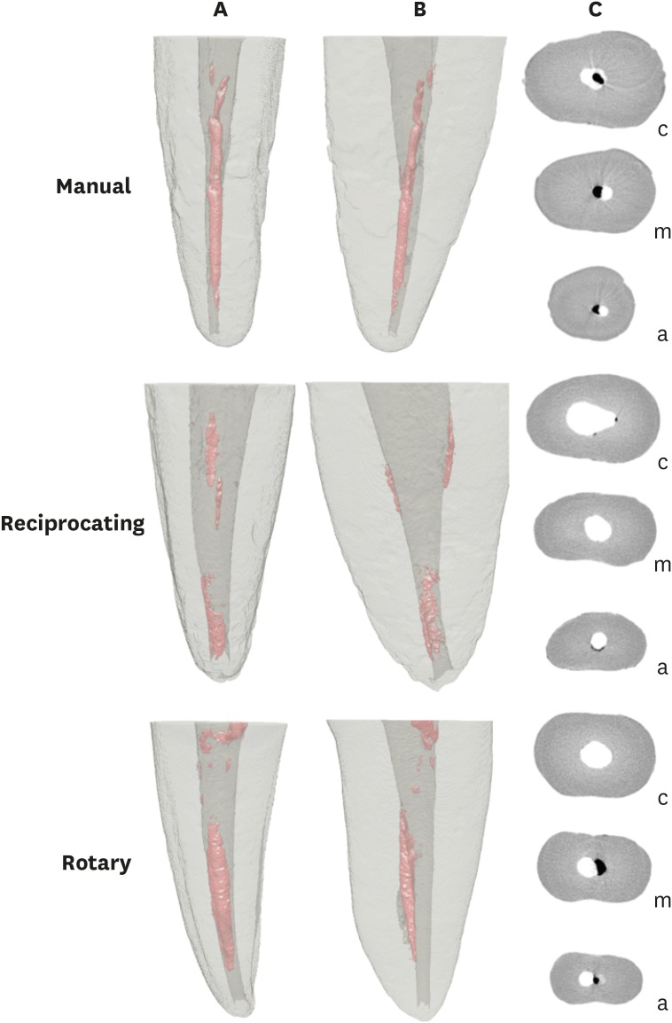
This study evaluated the amount of remaining root canal filling materials after retreatment procedures performed by undergraduate students using manual, rotary, and reciprocating techniques through micro-computed tomographic analysis. The incidence of instrument fracture and the instrumentation time were also evaluated. Materials and MethodsThirty maxillary single rooted teeth were prepared with Reciproc R25 files and filled with gutta-percha and AH Plus sealer by the continuous wave of condensation technique. Then, the specimens were assigned to 3 groups (n = 10), according to the retreatment technique used: manual, rotary, and reciprocating groups, which used K-file, Mtwo retreatment file, and Reciproc file, respectively. Retreatments were performed by undergraduate students. The sample was scanned after root canal filling and retreatment procedures, and the images of the canals were examined to quantify the amount of remaining filling material. The incidence of instrument fracture and the instrumentation time were recorded. ResultsRemaining filling material was observed in all specimens regardless of the technique used. The mean volume of remaining material was significantly lower in the Reciproc group than in the manual K-file and Mtwo retreatment groups (p < 0.05). The time required to achieve a satisfactory removal of canal filling material and refinement was significantly lower in the Mtwo retreatment and Reciproc groups (p < 0.05) when compared to the manual K-file group. No instrument fracture was observed in any of the groups. ConclusionsReciproc was the most effective instrument in the removal of canal fillings after retreatments performed by undergraduate students.
-
Citations
Citations to this article as recorded by  - Assessment of isthmus filling using two obturation techniques performed by students with different levels of clinical experience
Yang Yu, Chong-Yang Yuan, Xing-Zhe Yin, Xiao-Yan Wang
Journal of Dental Sciences.2024; 19(1): 169. CrossRef - Optical microscopy evaluation of root canal filling removal by beginner operators in posterior teeth
Bogdan Dimitriu, Ioana Suciu, Oana Elena Amza, Mihai Ciocârdel, Dana Bodnar, Ana Maria Cristina Țâncu, Mihaela Tanase, Maria Sabina Branescu, Mihaela Chirilă
Journal of Medicine and Life.2024; 17(6): 555. CrossRef - Micro-CT Study on the Supplementary Effect of XP-Endo Finisher R after Endodontic Retreatment with Mtwo-R
I Tsenova-Ilieva, V Dogandzhiyska, M Raykovska, E Karova
Nigerian Journal of Clinical Practice.2023; 26(12): 1844. CrossRef - Critical analysis of research methods and experimental models to study removal of root filling materials
Mahdi A. Ajina, Pratik K. Shah, Bun San Chong
International Endodontic Journal.2022; 55(S1): 119. CrossRef - Efficiency of Supplementary Contemporary Single-file Systems in Removing Filling Remnants from Oval-shaped Canals: An In Vitro Study
Neveen A Shaheen, Dalia A Sherif, Nahla G Elhelbawy
The Journal of Contemporary Dental Practice.2021; 22(9): 1055. CrossRef - Efficacy of an arrow‐shaped ultrasonic tip for the removal of residual root canal filling materials
Emmanuel J.N.L. Silva, Carolina O. de Lima, Ana F.A. Barbosa, Cláudio M. Ferreira, Bruno M. Crozeta, Ricardo T. Lopes
Australian Endodontic Journal.2021; 47(3): 467. CrossRef - XP‐endo Finisher R instrument optimizes the removal of root filling remnants in oval‐shaped canals
G. De‐Deus, F. G. Belladonna, A. S. Zuolo, D. M. Cavalcante, J. C. A. Carvalhal, M. Simões‐Carvalho, E. M. Souza, R. T. Lopes, E. J. N. L. Silva
International Endodontic Journal.2019; 52(6): 899. CrossRef
-
1,878
View
-
13
Download
-
7
Crossref
|