-
Micro-computed tomographic evaluation of canal retreatments performed by undergraduate students using different techniques
-
Emmanuel João Nogueira Leal Silva, Felipe Gonçalves Belladonna, Marianna Fernandes Carapiá, Brenda Leite Muniz, Mariana Santoro Rocha, Edson Jorge Lima Moreira
-
Restor Dent Endod 2018;43(1):e5. Published online January 15, 2018
-
DOI: https://doi.org/10.5395/rde.2018.43.e5
-
-
 Abstract Abstract
 PDF PDF PubReader PubReader ePub ePub
- Objectives
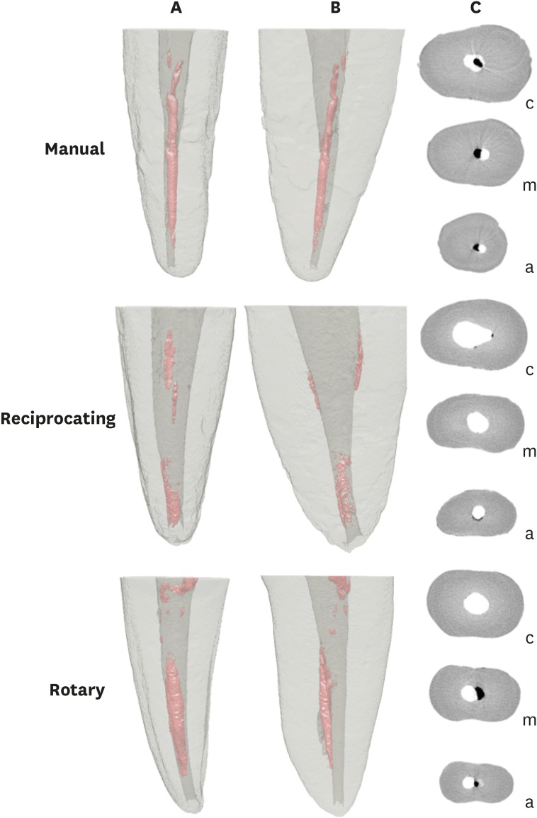
This study evaluated the amount of remaining root canal filling materials after retreatment procedures performed by undergraduate students using manual, rotary, and reciprocating techniques through micro-computed tomographic analysis. The incidence of instrument fracture and the instrumentation time were also evaluated. Materials and MethodsThirty maxillary single rooted teeth were prepared with Reciproc R25 files and filled with gutta-percha and AH Plus sealer by the continuous wave of condensation technique. Then, the specimens were assigned to 3 groups (n = 10), according to the retreatment technique used: manual, rotary, and reciprocating groups, which used K-file, Mtwo retreatment file, and Reciproc file, respectively. Retreatments were performed by undergraduate students. The sample was scanned after root canal filling and retreatment procedures, and the images of the canals were examined to quantify the amount of remaining filling material. The incidence of instrument fracture and the instrumentation time were recorded. ResultsRemaining filling material was observed in all specimens regardless of the technique used. The mean volume of remaining material was significantly lower in the Reciproc group than in the manual K-file and Mtwo retreatment groups (p < 0.05). The time required to achieve a satisfactory removal of canal filling material and refinement was significantly lower in the Mtwo retreatment and Reciproc groups (p < 0.05) when compared to the manual K-file group. No instrument fracture was observed in any of the groups. ConclusionsReciproc was the most effective instrument in the removal of canal fillings after retreatments performed by undergraduate students.
-
Citations
Citations to this article as recorded by  - Assessment of isthmus filling using two obturation techniques performed by students with different levels of clinical experience
Yang Yu, Chong-Yang Yuan, Xing-Zhe Yin, Xiao-Yan Wang
Journal of Dental Sciences.2024; 19(1): 169. CrossRef - Optical microscopy evaluation of root canal filling removal by beginner operators in posterior teeth
Bogdan Dimitriu, Ioana Suciu, Oana Elena Amza, Mihai Ciocârdel, Dana Bodnar, Ana Maria Cristina Țâncu, Mihaela Tanase, Maria Sabina Branescu, Mihaela Chirilă
Journal of Medicine and Life.2024; 17(6): 555. CrossRef - Micro-CT Study on the Supplementary Effect of XP-Endo Finisher R after Endodontic Retreatment with Mtwo-R
I Tsenova-Ilieva, V Dogandzhiyska, M Raykovska, E Karova
Nigerian Journal of Clinical Practice.2023; 26(12): 1844. CrossRef - Critical analysis of research methods and experimental models to study removal of root filling materials
Mahdi A. Ajina, Pratik K. Shah, Bun San Chong
International Endodontic Journal.2022; 55(S1): 119. CrossRef - Efficiency of Supplementary Contemporary Single-file Systems in Removing Filling Remnants from Oval-shaped Canals: An In Vitro Study
Neveen A Shaheen, Dalia A Sherif, Nahla G Elhelbawy
The Journal of Contemporary Dental Practice.2021; 22(9): 1055. CrossRef - Efficacy of an arrow‐shaped ultrasonic tip for the removal of residual root canal filling materials
Emmanuel J.N.L. Silva, Carolina O. de Lima, Ana F.A. Barbosa, Cláudio M. Ferreira, Bruno M. Crozeta, Ricardo T. Lopes
Australian Endodontic Journal.2021; 47(3): 467. CrossRef - XP‐endo Finisher R instrument optimizes the removal of root filling remnants in oval‐shaped canals
G. De‐Deus, F. G. Belladonna, A. S. Zuolo, D. M. Cavalcante, J. C. A. Carvalhal, M. Simões‐Carvalho, E. M. Souza, R. T. Lopes, E. J. N. L. Silva
International Endodontic Journal.2019; 52(6): 899. CrossRef
-
1,298
View
-
12
Download
-
7
Crossref
-
Comparison of canal transportation in simulated curved canals prepared with ProTaper Universal and ProTaper Gold systems
-
Emmanuel João Nogueira Leal Silva, Brenda Leite Muniz, Frederico Pires, Felipe Gonçalves Belladonna, Aline Almeida Neves, Erick Miranda Souza, Gustavo De-Deus
-
Restor Dent Endod 2016;41(1):1-5. Published online February 4, 2016
-
DOI: https://doi.org/10.5395/rde.2016.41.1.1
-
-
 Abstract Abstract
 PDF PDF PubReader PubReader ePub ePub
- Objectives
The purpose of this study was to assess the ability of ProTaper Gold (PTG, Dentsply Maillefer) in maintaining the original profile of root canal anatomy. For that, ProTaper Universal (PTU, Dentsply Maillefer) was used as reference techniques for comparison. Materials and MethodsTwenty simulated curved canals manufactured in clear resin blocks were randomly assigned to 2 groups (n = 10) according to the system used for canal instrumentation: PTU and PTG groups, upto F2 files (25/0.08). Color stereomicroscopic images from each block were taken exactly at the same position before and after instrumentation. All image processing and data analysis were performed with an open source program (FIJI). Evaluation of canal transportation was obtained for two independent canal regions: straight and curved levels. Student's t test was used with a cut-off for significance set at α = 5%. ResultsInstrumentation systems significantly influenced canal transportation (p < 0.0001). A significant interaction between instrumentation system and root canal level (p < 0.0001) was found. PTU and PTG systems produced similar canal transportation at the straight part, while PTG system resulted in lower canal transportation than PTU system at the curved part. Canal transportation was higher at the curved canal portion (p < 0.0001). ConclusionsPTG system produced overall less canal transportation in the curved portion when compared to PTU system.
-
Citations
Citations to this article as recorded by  - Shaping, and disinfecting abilities of ProTaper Universal, ProTaper Gold, and Twisted Files: A correlative microcomputed tomographic and bacteriologic analysis
Malavika Sivakumar, Ruchika Roongta Nawal, Sangeeta Talwar, CP Baveja, Rega Kumar, Sudha Yadav, S Santosh Kumar
Endodontology.2023; 35(1): 54. CrossRef - Advancing Nitinol: From heat treatment to surface functionalization for nickel–titanium (NiTi) instruments in endodontics
Wai-Sze Chan, Karan Gulati, Ove A. Peters
Bioactive Materials.2023; 22: 91. CrossRef - Comparative Evaluation of Root Canal Centering Ability of Two Heat-treated Single-shaping NiTi Rotary Instruments in Simulated Curved Canals: An In Vitro Study
Preethi Varadan, Chakravarthy Arumugam, Athira Shaji, R R Mathan
World Journal of Dentistry.2023; 14(6): 535. CrossRef - An Appraisal on Newer Endodontic File Systems: A Narrative Review
Shalini Singh, Kailash Attur, Anjali Oak, Mohammed Mustafa, Kamal Kumar Bagda, Nishtha Kathiria
The Journal of Contemporary Dental Practice.2023; 23(9): 944. CrossRef - Shaping ability of modern Nickel–Titanium rotary systems on the preparation of printed mandibular molars
Seda Falakaloglu, Emmanuel Silva, Burcu Topal, Emre İriboz, Mustafa Gündoğar
Journal of Conservative Dentistry.2022; 25(5): 498. CrossRef - An Investigation of the Accuracy and Reproducibility of 3D Printed Transparent Endodontic Blocks
Martin Smutný, Martin Kopeček, Aleš Bezrouk
Acta Medica (Hradec Kralove, Czech Republic).2022; 65(2): 59. CrossRef - Nitinol Type Alloys General Characteristics and Applications in Endodontics
Leszek A. Dobrzański, Lech B. Dobrzański, Anna D. Dobrzańska-Danikiewicz, Joanna Dobrzańska
Processes.2022; 10(1): 101. CrossRef - Impact of Endodontic Kinematics on Stress Distribution During Root Canal Treatment: Analysis of Photoelastic Stress
Shelyn Akari Yamakami, Julia Adornes Gallas, Igor Bassi Ferreira Petean, Aline Evangelista Souza-Gabriel, Manoel Sousa-Neto, Ana Paula Macedo, Regina Guenka Palma-Dibb
Journal of Endodontics.2022; 48(2): 255. CrossRef - Shaping ability of ProTaper Gold and WaveOne Gold nickel-titanium rotary instruments in simulated S-shaped root canals
Lu Shi, Junling Zhou, Jie Wan, Yunfei Yang
Journal of Dental Sciences.2022; 17(1): 430. CrossRef - A Comparative Study of Two Martensitic Alloy Systems in Endodontic Files Carried out by Unskilled Hands
Juan Algar, Alejandra Loring-Castillo, Ruth Pérez-Alfayate, Carmen Martín Carreras-Presas, Ana Suárez
Applied Sciences.2022; 12(12): 6289. CrossRef - Quantitative evaluation of apically extruded debris using TRUShape, TruNatomy, and WaveOne Gold in curved canals
Nehal Nabil Roshdy, Reham Hassan
BDJ Open.2022;[Epub] CrossRef - Comparison of Canal Transportation, Separation Rate, and Preparation Time between One Shape and Neoniti (Neolix): An In Vitro CBCT Study
Maryam Kuzekanani, Faranak Sadeghi, Nima Hatami, Maryam Rad, Mansoureh Darijani, Laurence James Walsh, Sivakumar Nuvvula
International Journal of Dentistry.2021; 2021: 1. CrossRef - Shaping ability of ProTaper Gold, One Curve, and Self-Adjusting File systems in severely curved canals: A cone-beam computed tomography study
MeenuG Singla, Hemanshi Kumar, Ritika Satija
Journal of Conservative Dentistry.2021; 24(3): 271. CrossRef - Cone-beam computed tomographic analysis of apical transportation and centering ratio of ProTaper and XP-endo Shaper NiTi rotary systems in curved canals: an in vitro study
Hamed Karkehabadi, Zeinab Siahvashi, Abbas Shokri, Nasrin Haji Hasani
BMC Oral Health.2021;[Epub] CrossRef - Mechanical Tests, Metallurgical Characterization, and Shaping Ability of Nickel-Titanium Rotary Instruments: A Multimethod Research
Emmanuel J.N.L. Silva, Jorge N.R. Martins, Carolina O. Lima, Victor T.L. Vieira, Francisco M. Braz Fernandes, Gustavo De-Deus, Marco A. Versiani
Journal of Endodontics.2020; 46(10): 1485. CrossRef - Micro-computed tomographic evaluation of a new system for root canal filling using calcium silicate-based root canal sealers
Mario Tanomaru-Filho, Fernanda Ferrari Esteves Torres, Jader Camilo Pinto, Airton Oliveira Santos-Junior, Karina Ines Medina Carita Tavares, Juliane Maria Guerreiro-Tanomaru
Restorative Dentistry & Endodontics.2020;[Epub] CrossRef - Comparison of vibration characteristics of file systems for root canal shaping according to file length
Seong-Jun Park, Se-Hee Park, Kyung-Mo Cho, Hyo-Jin Ji, Eun-Hye Lee, Jin-Woo Kim
Restorative Dentistry & Endodontics.2020;[Epub] CrossRef - New thermomechanically treated NiTi alloys – a review
J. Zupanc, N. Vahdat‐Pajouh, E. Schäfer
International Endodontic Journal.2018; 51(10): 1088. CrossRef - Shaping ability of four root canal instrumentation systems in simulated 3D-printed root canal models
David Christofzik, Andreas Bartols, Mahmoud Khaled Faheem, Doreen Schroeter, Birte Groessner-Schreiber, Christof E. Doerfer, Cyril Charles
PLOS ONE.2018; 13(8): e0201129. CrossRef - OPEN-SOURCE SOFTWARE IN DENTISTRY: A SYSTEMATIC REVIEW
Małgorzata Chruściel-Nogalska, Tomasz Smektała, Marcin Tutak, Katarzyna Sporniak-Tutak, Raphael Olszewski
International Journal of Technology Assessment in Health Care.2017; 33(4): 487. CrossRef - Mechanical Properties of Various Heat-treated Nickel-titanium Rotary Instruments
Hye-Jin Goo, Sang Won Kwak, Jung-Hong Ha, Eugenio Pedullà, Hyeon-Cheol Kim
Journal of Endodontics.2017; 43(11): 1872. CrossRef - A comparison of the shaping ability of three nickel-titanium rotary instruments: a micro-computed tomography study via a contrast radiopaque technique in vitro
Zhao Wei, Zhi Cui, Ping Yan, Han Jiang
BMC Oral Health.2017;[Epub] CrossRef - Root Canal Transportation and Centering Ability of Nickel-Titanium Rotary Instruments in Mandibular Premolars Assessed Using Cone-Beam Computed Tomography
Iussif Mamede-Neto, Alvaro Henrique Borges, Orlando Aguirre Guedes, Durvalino de Oliveira, Fábio Luis Miranda Pedro, Carlos Estrela
The Open Dentistry Journal.2017; 11(1): 71. CrossRef - Blue Thermomechanical Treatment Optimizes Fatigue Resistance and Flexibility of the Reciproc Files
Gustavo De-Deus, Emmanuel João Nogueira Leal Silva, Victor Talarico Leal Vieira, Felipe Gonçalves Belladonna, Carlos Nelson Elias, Gianluca Plotino, Nicola Maria Grande
Journal of Endodontics.2017; 43(3): 462. CrossRef
-
1,608
View
-
7
Download
-
24
Crossref
|


