-
Porosity and pore size distribution in high-viscosity and conventional glass ionomer cements: a micro-computed tomography study
-
Aline Borburema Neves, Laísa Inara Gracindo Lopes, Tamiris Gomes Bergstrom, Aline Saddock Sá da Silva, Ricardo Tadeu Lopes, Aline de Almeida Neves
-
Restor Dent Endod 2021;46(4):e57. Published online October 29, 2021
-
DOI: https://doi.org/10.5395/rde.2021.46.e57
-
-
 Abstract Abstract
 PDF PDF PubReader PubReader ePub ePub
- Objectives
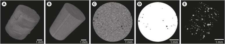
This study aimed to compare and evaluate the porosity and pore size distribution of high-viscosity glass ionomer cements (HVGICs) and conventional glass ionomer cements (GICs) using micro-computed tomography (micro-CT). Materials and MethodsForty cylindrical specimens (n = 10) were produced in standardized molds using HVGICs and conventional GICs (Ketac Molar Easymix, Vitro Molar, MaxxionR, and Riva Self-Cure). The specimens were prepared according to ISO 9917-1 standards, scanned in a high-energy micro-CT device, and reconstructed using specific parameters. After reconstruction, segmentation procedures, and image analysis, total porosity and pore size distribution were obtained for specimens in each group. After checking the normality of the data distribution, the Kruskal-Wallis test followed by the Student-Newman-Keuls test was used to detect differences in porosity among the experimental groups with a 5% significance level. ResultsKetac Molar Easymix showed statistically significantly lower total porosity (0.15%) than MaxxionR (0.62%), Riva (0.42%), and Vitro Molar (0.57%). The pore size in all experimental cements was within the small-size range (< 0.01 mm3), but Vitro Molar showed statistically significantly more pores/defects with a larger size (> 0.01 mm3). ConclusionsMajor differences in porosity and pore size were identified among the evaluated GICs. Among these, the Ketac Molar Easymix HVGIC showed the lowest porosity and void size.
-
Citations
Citations to this article as recorded by  - The effect of contouring instruments on immediate quality and porosity of direct restorations
Carlos Soler-Tornero, Pekka Toivonen, Jaakko Suorsa, Sakari S. Karhula, Simo Saarakkala, Vuokko Anttonen, Jukka Leinonen
Clinical Oral Investigations.2025;[Epub] CrossRef - Impact of spacers and thermocycling on porosity and gaps in class II endodontic temporary restorations evaluated by microcomputed tomography
Fahda N. Algahtani, Manal Alkadi, Hiba R. Talic, Sarah S. AlShalawi, Lujain M. Alqarni, Reem M. Barakat, Rasha Haridy, Sara M. ElKhateeb, Rahaf A. Almohareb
Scientific Reports.2025;[Epub] CrossRef - Influence of Human Blood Contamination on Microhardness of Glass-Ionomer Cements and Glass-Hybrid Material
Katarina Franić, Ana Brundić, Jurica Matijević, Ana Ivanišević, Ivana Miletić, Anja Baraba
Materials.2025; 18(17): 4075. CrossRef - Effect of crown seating methods on the remnant cement in the subgingival region of a cement-retained implant crown
Fanghui Ji, Ji Suk Shim, Jeongyol Lee, Hwiseong Oh, Jae Jun Ryu
Scientific Reports.2024;[Epub] CrossRef - Enhancing Wear Resistance in Glass Ionomer Cement through Green-mediated Chitosan-, Titanium-, Zirconium-, and Hydroxyapatite-based Nanocomposites: An Analysis before and after Chewing Simulator Endurance
Srinavasa Surya Sitaram, Jessy Paulraj, Subhabrata Maiti, Rajeshkumar Shanmugam
International Journal of Clinical Pediatric Dentistry.2024; 17(11): 1229. CrossRef - The effect of mesoporous silica doped with silver nanoparticles on glass ionomer cements; physiochemical, mechanical and ion release analysis
Syed Saad Bin Qasim, Ali Bmuajdad
BMC Oral Health.2024;[Epub] CrossRef - Hyperbaric Pressure Effect on Dental Luting Cements
Secil OZKAN ATA, Nazım ATA, Rıfat UGURLUTAN
Journal of Basic and Clinical Health Sciences.2023; 7(1): 464. CrossRef - In Vitro Comparison of Differences in Setting Time of Premixed Calcium Silicate-Based Mineral Trioxide Aggregate According to Moisture Content of Gypsum
Hyun-Jin Kim, Jun-Seok Lee, Dong-Hoon Gwak, Yong-Seok Ko, Chun-Il Lim, Seung-Youl Lee
Materials.2023; 17(1): 35. CrossRef - Adhesion and Surface Roughness of Apatite-Containing Carbomer and Improved Ionically Bioactive Resin Compared to Glass Ionomers
Handan Yıldırım Işık, Aylin Çilingir
Journal of Functional Biomaterials.2023; 14(7): 367. CrossRef - An influence of finishing procedures and protective coating on the ultrastructure of conventional and hybrid glass ionomer cement restorations
Antonije Stankovic, Jelena Popovic, Marija Nikolic, Aleksandar Mitic, Nenad Stosic, Radomir Barac, Aleksandra Milovanovic
Stomatoloski glasnik Srbije.2023; 70(3): 138. CrossRef - Effect of aging on mechanical and antibacterial properties of fluorinated graphene reinforced glass ionomer: In vitro study
Suzan Khaled Arafa, Dalia Ibrahim Sherief, Mohamed Salah Nassif
Journal of the Mechanical Behavior of Biomedical Materials.2023; 142: 105803. CrossRef
-
3,214
View
-
18
Download
-
8
Web of Science
-
11
Crossref
-
Push-out bond strength of a self-adhesive resin cement used as endodontic sealer
-
Eduardo Diogo Gurgel-Filho, Felipe Coelho Lima, Vicente de Paula Aragão Saboia, Tauby de Souza Coutinho-Filho, Aline de Almeida Neves, Emmanuel João Nogueira Leal da Silva
-
Restor Dent Endod 2014;39(4):282-287. Published online August 20, 2014
-
DOI: https://doi.org/10.5395/rde.2014.39.4.282
-
-
Abstract
PDFPubReaderePub
- Objectives
The aim of the present study was to investigate the bond strength of RelyX Unicem (3M) to root canal dentin when used as an endodontic sealer. Materials and MethodsSamples of 24 single-rooted teeth were prepared with Gates Glidden drills and K3 files. After that, the roots were randomly assigned to three experimental groups (n = 8) according to the filling material, (1) AH Plus (Dentsply De Trey GmbH)/Gutta-Percha cone; (2) Epiphany SE (Pentron)/Resilon cone; (3) RelyX Unicem/Gutta-Percha cone. All roots were filled using a single cone technique associated to vertical condensation. After the filling procedures, each tooth was prepared for a push-out bond strenght test by cutting 1 mm-thick root slices. Loading was performed on a universal testing machine at a speed of 0.5 mm/min. One-way analysis of variance and Tukey test for multiple comparisons were used to compare the results among the experimental groups. ResultsEpiphany SE/Resilon showed significantly lower push-out bond strength than both AH Plus/Gutta-Percha and RelyX Unicem/Gutta-Percha (p < 0.05). There was no significant difference in bond strength between AH Plus/Gutta-Percha and RelyX Unicem/Gutta-Percha (p > 0.05). ConclusionsUnder the present in vitro conditions, bond strength to root dentin promoted by RelyX Unicem was similar to AH Plus. Epiphany SE/Resilon resulted in lower bond strength values when compared to both materials.
-
Citations
Citations to this article as recorded by - In vitro comparative evaluation of physicochemical and mechanical properties, cytocompatibility, and antimicrobial efficacy of various bioceramic root canal sealers
Fushi Wang, Jiaxing Li, Jingjing Wan, Siyuan Li, Shijia Tang, Li Wang, Liuyan Meng
Ceramics International.2026; 52(7): 9561. CrossRef - In-Vitro Comparative Adhesion Evaluation of Bioceramic and Dual-Cure Resin Endodontic Sealers Using SEM, AFM, Push-Out and FTIR
Radu Marcel Chisnoiu, Marioara Moldovan, Doina Prodan, Andrea Maria Chisnoiu, Dana Hrab, Ada Gabriela Delean, Alexandrina Muntean, Doina Iulia Rotaru, Ovidiu Pastrav, Mihaela Pastrav
Applied Sciences.2021; 11(10): 4454. CrossRef - Push-out Bond Strength of Fiber Posts Cemented Using New Universal Adhesives on Etched and Nonetched Intraradicular Dentin
Hani F Ounsi, Simone Grandini, Marco Ferrari, Valentina Spicciarelli, Giacomo Corsentino, Crystal Marruganti
The Journal of Contemporary Dental Practice.2020; 21(1): 91. CrossRef - Comparison of push-out bond strength of three different obturating systems to intraradicular dentin: An In vitro study
MohammedKhwaja Moinuddin, LKarthik Prasad, Nimeshika Ramachandruni, Shekar Kamishetty, RaviChandra Cherkupalli
Contemporary Clinical Dentistry.2019; 10(4): 631. CrossRef - The influence of methodological variables on the push‐out resistance to dislodgement of root filling materials: a meta‐regression analysis
F. M. Collares, F. F. Portella, S. B. Rodrigues, R. K. Celeste, V. C. B. Leitune, S. M. W. Samuel
International Endodontic Journal.2016; 49(9): 836. CrossRef - Effect of photon induced photoacoustic streaming (PIPS) on bond strength to dentine of two root canal filling materials
Ivana Miletić, Nicoletta Chieffi, Carlo Rengo, Marco Ferrari, Dan Nathanson, Anja Baraba
Lasers in Surgery and Medicine.2016; 48(10): 951. CrossRef
-
2,244
View
-
6
Download
-
6
Crossref
|