-
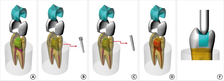
Effect of the restorative technique on load-bearing capacity, cusp deflection, and stress distribution of endodontically-treated premolars with MOD restoration
-
Daniel Maranha da Rocha, João Paulo Mendes Tribst, Pietro Ausiello, Amanda Maria de Oliveira Dal Piva, Milena Cerqueira da Rocha, Rebeca Di Nicoló, Alexandre Luiz Souto Borges
-
Restor Dent Endod 2019;44(3):e33. Published online August 7, 2019
-
DOI: https://doi.org/10.5395/rde.2019.44.e33
-
-
 Abstract Abstract
 PDF PDF PubReader PubReader ePub ePub
- Objectives
To evaluate the influence of the restorative technique on the mechanical response of endodontically-treated upper premolars with mesio-occluso-distal (MOD) cavity. Materials and MethodsForty-eight premolars received MOD preparation (4 groups, n = 12) with different restorative techniques: glass ionomer cement + composite resin (the GIC group), a metallic post + composite resin (the MP group), a fiberglass post + composite resin (the FGP group), or no endodontic treatment + restoration with composite resin (the CR group). Cusp strain and load-bearing capacity were evaluated. One-way analysis of variance and the Tukey test were used with α = 5%. Finite element analysis (FEA) was used to calculate displacement and tensile stress for the teeth and restorations. ResultsMP showed the highest cusp (p = 0.027) deflection (24.28 ± 5.09 µm/µm), followed by FGP (20.61 ± 5.05 µm/µm), CR (17.72 ± 6.32 µm/µm), and GIC (17.62 ± 7.00 µm/µm). For load-bearing, CR (38.89 ± 3.24 N) showed the highest, followed by GIC (37.51 ± 6.69 N), FGP (29.80 ± 10.03 N), and MP (18.41 ± 4.15 N) (p = 0.001) value. FEA showed similar behavior in the restorations in all groups, while MP showed the highest stress concentration in the tooth and post. ConclusionsThere is no mechanical advantage in using intraradicular posts for endodontically-treated premolars requiring MOD restoration. Filling the pulp chamber with GIC and restoring the tooth with only CR showed the most promising results for cusp deflection, failure load, and stress distribution.
-
Citations
Citations to this article as recorded by  - How to adaptively balance ‘classic’ or ‘conservative’ approaches in tooth defect management: a 3D-finite element analysis study
Jiani Xu, Xu Liang, Lili Hu, Chen Sun, Zhipeng Zhang, Jiawei Yang, Jie Wang
BMC Oral Health.2025;[Epub] CrossRef - Inkjet-printed strain gauge sensors: Materials, manufacturing, and emerging applications
Lara Abdel Salam, Samir Mustapha, Alexandra Mikhael, Nisrine Bakri, Sahera Saleh, Massoud L. Khraiche
Sensors and Actuators A: Physical.2025; 394: 116934. CrossRef - Influence of endodontic access cavity design on mechanical properties of a first mandibular premolar tooth: a finite element analysis study
Taha Özyürek, Gülşah Uslu, Burçin Arıcan, Mustafa Gündoğar, Mohammad Hossein Nekoofar, Paul Michael Howell Dummer
Clinical Oral Investigations.2024;[Epub] CrossRef - Comparison of the Effect of Different Cavity Designs and Temporary Restoration Materials on the Fracture Resistance of Upper Premolars, Undergone Re-treatment: An In-Vitro Study
Parnian Alavinejad, Mohammad Yazdizadeh, Ali Mombeinipour, Ebrahim Karimzadeh
Proceedings of the National Academy of Sciences, India Section B: Biological Sciences.2024; 94(3): 677. CrossRef - Fracture resistance and failure mode of endodontically treated premolars reconstructed by different preparation approaches: Cervical margin relocation and crown lengthening with complete and partial ferrule with three different post and core systems
Mehran Falahchai, Naghmeh Musapoor, Soroosh Mokhtari, Yasamin Babaee Hemmati, Hamid Neshandar Asli
Journal of Prosthodontics.2024; 33(8): 774. CrossRef - Comparison of the stress distribution in base materials and thicknesses in composite resin restorations
Min-Kwan Jung, Mi-Jeong Jeon, Jae-Hoon Kim, Sung-Ae Son, Jeong-Kil Park, Deog-Gyu Seo
Heliyon.2024; 10(3): e25040. CrossRef -
Fracture resistance and failure pattern of endodontically treated maxillary premolars restored with transfixed glass fiber post: an
in vitro
and finite element analysis
Saleem Abdulrab, Greta Geerts, Ganesh Thiagarajan
Computer Methods in Biomechanics and Biomedical Engineering.2024; 27(4): 419. CrossRef - Influence of size-anatomy of the maxillary central incisor on the biomechanical performance of post-and-core restoration with different ferrule heights
Domingo Santos Pantaleón, João Paulo Mendes Tribst, Franklin García-Godoy
The Journal of Advanced Prosthodontics.2024; 16(2): 77. CrossRef - Influence of internal angle and shape of the lining on residual stress of Class II molar restorations
Qianqian Zuo, Annan Li, Haidong Teng, Zhan Liu
Computer Methods in Biomechanics and Biomedical Engineering.2024; 27(5): 680. CrossRef - Evaluation of stress distribution in coronal base and restorative materials: A narrative review of finite element analysis studies
Yelda Polat, İzzet Yavuz
Conservative Dentistry Journal.2024; 14(2): 47. CrossRef - The influence of horizontal glass fiber posts on fracture strength and fracture pattern of endodontically treated teeth: A systematic review and meta‐analysis of in vitro studies
Saleem Abdulrab, Greta Geerts, Sadeq Ali Al‐Maweri, Mohammed Nasser Alhajj, Hatem Alhadainy, Raidan Ba‐Hattab
Journal of Prosthodontics.2023; 32(6): 469. CrossRef - Stress distribution of a novel bundle fiber post with curved roots and oval canals
Deniz Yanık, Nurullah Turker
Journal of Esthetic and Restorative Dentistry.2022; 34(3): 550. CrossRef - The Effect of Endodontic Treatment and Thermocycling on Cuspal Deflection of Teeth Restored with Different Direct Resin Composites
Cansu Atalay, Ayse Ruya Yazici, Aynur Sidika Horuztepe, Emre Nagas
Conservative Dentistry and Endodontic Journal.2022; 6(2): 38. CrossRef - The use of different adhesive filling material and mass combinations to restore class II cavities under loading and shrinkage effects: a 3D-FEA
P. Ausiello, S. Ciaramella, A. De Benedictis, A. Lanzotti, J. P. M. Tribst, D. C. Watts
Computer Methods in Biomechanics and Biomedical Engineering.2021; 24(5): 485. CrossRef - Biomechanical Analysis of a Custom-Made Mouthguard Reinforced With Different Elastic Modulus Laminates During a Simulated Maxillofacial Trauma
João Paulo Mendes Tribst, Amanda Maria de Oliveira Dal Piva, Pietro Ausiello, Arianna De Benedictis, Marco Antonio Bottino, Alexandre Luiz Souto Borges
Craniomaxillofacial Trauma & Reconstruction.2021; 14(3): 254. CrossRef - Mechanical Assessment of Glass Ionomer Cements Incorporated with Multi-Walled Carbon Nanotubes for Dental Applications
Manuela Spinola, Amanda Maria Oliveira Dal Piva, Patrícia Uchôas Barbosa, Carlos Rocha Gomes Torres, Eduardo Bresciani
Oral.2021; 1(3): 190. CrossRef - Stress Concentration of Endodontically Treated Molars Restored with Transfixed Glass Fiber Post: 3D-Finite Element Analysis
Alexandre Luiz Souto Borges, Manassés Tercio Vieira Grangeiro, Guilherme Schmitt de Andrade, Renata Marques de Melo, Kusai Baroudi, Laís Regiane Silva-Concilio, João Paulo Mendes Tribst
Materials.2021; 14(15): 4249. CrossRef - Computer Aided Design Modelling and Finite Element Analysis of Premolar Proximal Cavities Restored with Resin Composites
Amanda Guedes Nogueira Matuda, Marcos Paulo Motta Silveira, Guilherme Schmitt de Andrade, Amanda Maria de Oliveira Dal Piva, João Paulo Mendes Tribst, Alexandre Luiz Souto Borges, Luca Testarelli, Gabriella Mosca, Pietro Ausiello
Materials.2021; 14(9): 2366. CrossRef - Effect of Shrinking and No Shrinking Dentine and Enamel Replacing Materials in Posterior Restoration: A 3D-FEA Study
Pietro Ausiello, Amanda Maria de Oliveira Dal Piva, Alexandre Luiz Souto Borges, Antonio Lanzotti, Fausto Zamparini, Ettore Epifania, João Paulo Mendes Tribst
Applied Sciences.2021; 11(5): 2215. CrossRef - Effect of Fiber-Reinforced Composite and Elastic Post on the Fracture Resistance of Premolars with Root Canal Treatment—An In Vitro Pilot Study
Jesús Mena-Álvarez, Rubén Agustín-Panadero, Alvaro Zubizarreta-Macho
Applied Sciences.2020; 10(21): 7616. CrossRef
-
2,011
View
-
21
Download
-
20
Crossref
-
Influence of thickness and incisal extension of indirect veneers on the biomechanical behavior of maxillary canine teeth
-
Victória Luswarghi Souza Costa, João Paulo Mendes Tribst, Eduardo Shigueyuki Uemura, Dayana Campanelli de Morais, Alexandre Luiz Souto Borges
-
Restor Dent Endod 2018;43(4):e48. Published online November 12, 2018
-
DOI: https://doi.org/10.5395/rde.2018.43.e48
-
-
 Abstract Abstract
 PDF PDF PubReader PubReader ePub ePub
- Objectives
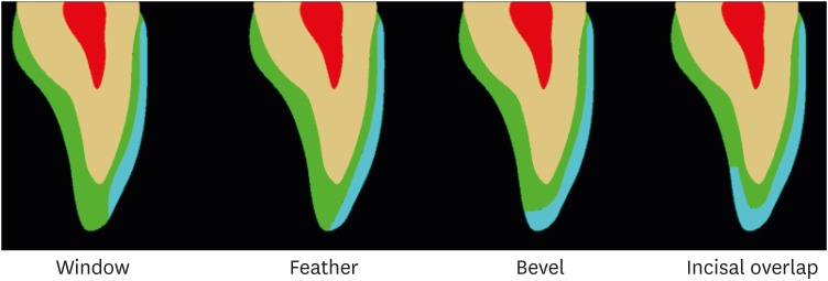
To analyze the influence of thickness and incisal extension of indirect veneers on the stress and strain generated in maxillary canine teeth. Materials and MethodsA 3-dimensional maxillary canine model was validated with an in vitro strain gauge and exported to computer-assisted engineering software. Materials were considered homogeneous, isotropic, and elastic. Each canine tooth was then subjected to a 0.3 and 0.8 mm reduction on the facial surface, in preparations with and without incisal covering, and restored with a lithium disilicate veneer. A 50 N load was applied at 45° to the long axis of the tooth, on the incisal third of the palatal surface of the crown. ResultsThe results showed a mean of 218.16 µstrain of stress in the in vitro experiment, and 210.63 µstrain in finite element analysis (FEA). The stress concentration on prepared teeth was higher at the palatal root surface, with a mean value of 11.02 MPa and varying less than 3% between the preparation designs. The veneers concentrated higher stresses at the incisal third of the facial surface, with a mean of 3.88 MPa and a 40% increase in less-thick veneers. The incisal cover generated a new stress concentration area, with values over 48.18 MPa. ConclusionsThe mathematical model for a maxillary canine tooth was validated using FEA. The thickness (0.3 or 0.8 mm) and the incisal covering showed no difference for the tooth structure. However, the incisal covering was harmful for the veneer, of which the greatest thickness was beneficial.
-
Citations
Citations to this article as recorded by 
-
1,826
View
-
14
Download
-
7
Crossref
|


