Articles
- Page Path
- HOME > Restor Dent Endod > Volume 28(3); 2003 > Article
- Original Article Color changes in composite resins exposed to xenon lamp
- Young-Gon Cho, Jeong-Il Seo, Soo-Mee Kim, Jin-Ho Jeong, Young-Gon Lee
-
2003;28(3):-202.
DOI: https://doi.org/10.5395/JKACD.2003.28.3.195
Published online: May 31, 2003
Department of Conservative Dentistry, College of dentistry, Chosun University, Korea.
- Corresponding author (ygcho@mail.chosun.ac.kr)
Copyright © 2003 Korean Academy of Conservative Dentistry
- 869 Views
- 1 Download
- 1 Crossref
Abstract
-
The purpose of this study was to evaluate the color changes of the composite resin resulting from xenon lamp exposure in different environments. Composite resin (Z 250 ; shade A1, A2, A3, A3.5, and A4) were applied in a cylindrical metal mold. Seventy five specimens according to environments of exposure were made as follows;Group I: aluminum foiling of the specimens in the air at 37℃ for 1 day and 7 days.Group II: exposure of xenon lamp to the specimens in the air at 37℃ for 1 day and 7 days.Group III: exposure of xenon lamp to the specimens in distilled water at 37℃ for 1 day and 7 days.The color characteristics (L*,a*,b*) of the specimens before and after exposure of xenon lamp were measured by spectrophotometer and the total color differences (ΔE*) were computed.The results obtained were as follows:
In all groups except A1 shade of group III, the ΔE* values presented below 2.0, and group III showed the highest ΔE* values followed by group II and group I in a decreasing order(p<0.05).
In all shades and groups, the more the exposure time of xenon lamp and the lighter the shade were, the higher the tendency for discoloration (p<0.05).
The composite resins which was exposed to xenon lamp in the distilled water was more discolored than those in the air (p<0.05).
The major changes of composite resins which were exposed to xenon lamp in the air were an increase in yellowness through a positive shift of the b* value, and those in the distilled water were an increase in darkness and yellowness through a negative shift of the L* value and a positive shift of the b* value.
- 1. Brauer GM. Color changes of composites on exposure to various energy sources. Dent Mater. 1988;4: 55-59.ArticlePubMed
- 2. Fruits TJ, Duncanson MG, Miranda FJ. In vitro weathering of selected direct esthetic reto-rative materials. Quintessence Int. 1997;28: 409-414.PubMed
- 3. Luce MS, Campbell CE. Stain potential of four microfilled composites. J Prosthet Dent. 1988;60: 151-154.PubMed
- 4. Powers JM, Fan PL, Raptis CN. Color stability of new composite restorative materials under accelerated aging. J Dent Res. 1980;59: 2071-2074.ArticlePubMedPDF
- 5. Stober T, Gilde H, Lenz P. Color stabiliy of highly filled composite resin materials for facings. Dent Mater. 2001;17: 87-94.PubMed
- 6. Shin HS, Hwang HK, Cho YG. A study on the staining tendency of esthetic restorative materials. J Korean Acad Conserv Dent. 1995;20: 372-383.
- 7. Chan KC, Fuller JL, Hormati AA. The ability of foods to stain two composite resin. J Prosthet Dent. 1980;43: 542-545.PubMed
- 8. Barkmeier WW, Cooley RL. Evaluation of surface finish of microfilled resins. J Esthet Dent. 1989;1: 139-143.ArticlePubMed
- 9. Burrow MF, Makinson OF. Color change in light-cured resins exposed to daylight. Quintessence Int. 1991;22: 447-452.PubMed
- 10. Kim MJ, Cho YG. Color Chances in Composites According to Various Light Curing Sources. J Korean Acad Conserv Dent. 2002;27: 87-94.
- 11. Ji CK. Lighting Theory. 2002;Seoul: Munundang; 80-81.
- 12. Leibrock A, Rosentritt M, Lang R, Behr M, Handel G. Colour stability of visible light-curing hybrid composites. Eur J Prosthodont Restor Dent. 1997;5: 125-130.PubMed
- 13. Plastics-methods of exposure to laboratory light. International Standard ISO 4892. 1981.
- 14. New American dental Association Specification No. 27 for direct filling resins. J Am Dent Assoc. 1977;94: 1191-1194.PubMed
- 15. Noie F, Keeefe KL, Powers JM. Color stability of resin cements after accelerated aging. Int J Prosthodont. 1995;8: 51-55.PubMed
- 16. Swift EJ Jr, Hammel SA, Lund PS. Colorimetric evaluatioin of vita shade resin composites. Int J Prosthodont. 1994;7: 356-361.PubMed
- 17. Asmussen E. Factor affecting the color stability of restorative resins. Acta Odontol Scand. 1983;41: 11-18.PubMed
- 18. Ferracane JL, Moser JB, Greener EH. Ultraviolet light-induced yellowing of dental resto-rative resins. J Prosthet Dent. 1985;54: 483-487.PubMed
- 19. Powers JM, Dennison JB, Koran A. Color stability of restorative resins under accelerated aging. J Dent Res. 1978;57: 964-970.PubMed
- 20. Uchida H, Vaidyanathan J, Viswanadhan T, Vaidyanathan TK. Color stability of dental composites as a function of shade. J Prosthet Dent. 1998;79: 372-377.PubMed
- 21. Hintze H. Klinische und Physikalische Untersuchungen der Farbbestandigkeit von Zahnarztlichen F llungskunststoffen. Freie Universität Berlin; Inaugural-Dissertation.
- 22. Viohl J. Color stability of dental resins. Quintessence Int. 1980;3: Report 1860.
- 23. Viohl J. Verfarbung von Kunststoffen durch unterschiedliche Lichtquellen. Dtsch Zahnarztl Z. 1976;31: 910-914.PubMed
- 24. Choi HK, Kang TE, Kim JH, Park HM. Lighting Installation and Design. 2000;Seoul: Sungandang; 82-83.
- 25. Asmussen E. An accelerated test for color stability of restorative resins. Acta Odontol Scand. 1981;39: 329-332.
- 26. Seghi RR, Jonston WM, O'Brien WJ. Performance assessment of colorimetric devices on dental porcelain. J Dent Res. 1989;68: 1755-1759.PubMed
- 27. Seghi RR, Gritz MD, Kim J. Colorimetric changes in composites resulting fromvisible-light-initiated polymerization. Dent Mater. 1990;6: 133-137.ArticlePubMed
- 28. Phillips RW. Skinner's science of dental materials. 1982;8th ed. Philadelphia: WB Saunders; 232.
REFERENCES
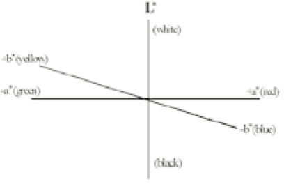
Tables & Figures
REFERENCES
Citations

- Effects of the color components of light-cured composite resin before and after polymerization on degree of conversion and flexural strength
Ji-A Yoo, Byeong-Hoon Cho
Journal of Korean Academy of Conservative Dentistry.2011; 36(4): 324. CrossRef

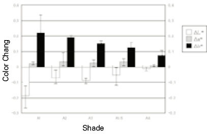
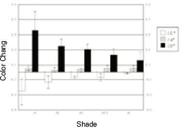
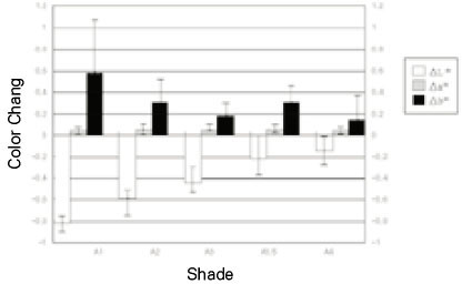
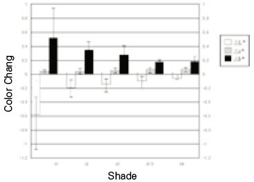
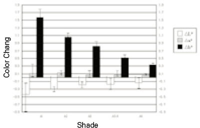
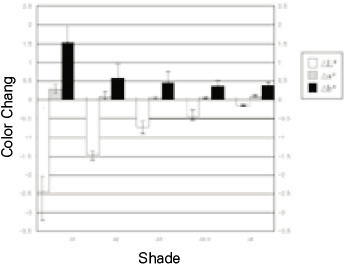
Fig. 1
Fig. 2
Fig. 3
Fig. 4
Fig. 5
Fig. 6
Fig. 7
Group classification.
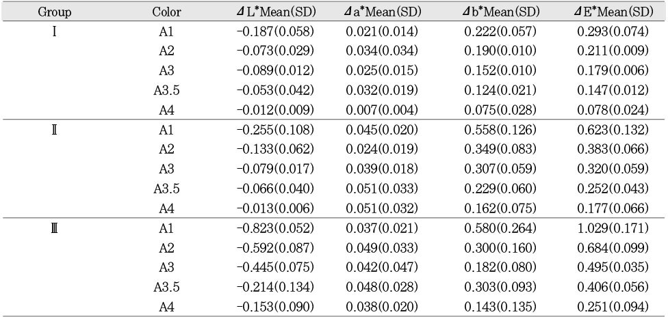
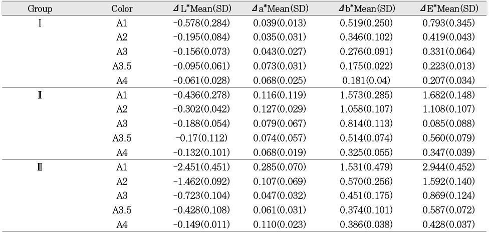
Result of color changes of group I, II and III after exposure of xenon lamp for 1 day.
ΔL*,Δa*,Δb*: color difference, ΔE*: total color difference.
Standard deviations are in parentheses.
Result of color changes of group I, II and III after exposure of xenon lamp for 7 days.
ΔL*,Δa*,Δb*: color difference, ΔE*: total color difference.
Standard deviations are in parentheses.
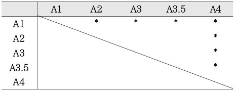
Statistical analysis of ΔE* of groupI between each shades by Tukey's HSD test (exposure to xenon lamp for 1 day).
*: significant differences (p<0.05)
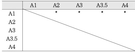
Statistical analysis of ΔE* of groupI between each shades by Turkey's HSD test (exposure to xenon lamp for 7 days).
*: significant differences (p<0.05)
ΔL*,Δa*,Δb*: color difference, ΔE*: total color difference. Standard deviations are in parentheses.
ΔL*,Δa*,Δb*: color difference, ΔE*: total color difference. Standard deviations are in parentheses.
*: significant differences (p<0.05)
*: significant differences (p<0.05)

 KACD
KACD


 ePub Link
 Cite
Cite

