Search
- Page Path
- HOME > Search
- Bonding of a resin-modified glass ionomer cement to dentin using universal adhesives
- Muhittin Ugurlu
- Restor Dent Endod 2020;45(3):e36. Published online June 15, 2020
- DOI: https://doi.org/10.5395/rde.2020.45.e36

-
 Abstract
 PDF
PDF PubReader
 ePub
ePub Objectives This study aims to assess the effect of universal adhesives pretreatment on the bond strength of resin-modified glass ionomer cement to dentin.
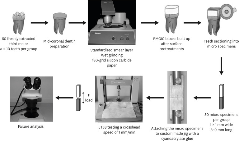
Materials and Methods Fifty caries-free human third molars were employed. The teeth were randomly assigned into five groups (
n = 10) based on dentin surface pretreatments: Single Bond Universal (3M Oral Care), Gluma Bond Universal (Heraeus Kulzer), Prime&Bond Elect (Dentsply), Cavity Conditioner (GC) and control (no surface treatment). After Fuji II LC (GC) was bonded to the dentin surfaces, the specimens were stored for 7 days at 37°C. The specimens were segmented into microspecimens, and the microspecimens were subjugated to microtensile bond strength testing (1.0 mm/min). The modes of failure analyzed using a stereomicroscope and scanning electron microscopy. Data were statistically analyzed with one-way analysis of variance and Duncan tests (p = 0.05).Results The surface pretreatments with the universal adhesives and conditioner increased the bond strength of Fuji II LC to dentin (
p < 0.05). Single Bond Universal and Gluma Bond Universal provided higher bond strength to Fuji II LC than Cavity Conditioner (p < 0.05). The bond strengths obtained from Prime&Bond Elect and Cavity Conditioner were not statistically different (p > 0.05).Conclusions The universal adhesives and polyacrylic acid conditioner could increase the bond strength of resin-modified glass ionomer cement (RMGIC) to dentin. The use of universal adhesives before the application of RMGIC may be more beneficial in improving bond strength.
- 31 View
- 0 Download

- Elemental analysis of caries-affected root dentin and artificially demineralized dentin
- Young-Hye Sung, Ho-Hyun Son, Keewook Yi, Juhea Chang
- Restor Dent Endod 2016;41(4):255-261. Published online August 19, 2016
- DOI: https://doi.org/10.5395/rde.2016.41.4.255
-
 Abstract
 PDF
PDF PubReader
 ePub
ePub Objectives This study aimed to analyze the mineral composition of naturally- and artificially-produced caries-affected root dentin and to determine the elemental incorporation of resin-modified glass ionomer (RMGI) into the demineralized dentin.
Materials and Methods Box-formed cavities were prepared on buccal and lingual root surfaces of sound human premolars (
n = 15). One cavity was exposed to a microbial caries model using a strain of Streptococcus mutans. The other cavity was subjected to a chemical model under pH cycling. Premolars and molars with root surface caries were used as a natural caries model (n = 15). Outer caries lesion was removed using a carbide bur and a hand excavator under a dyeing technique and restored with RMGI (FujiII LC, GC Corp.). The weight percentages of calcium (Ca), phosphate (P), and strontium (Sr) and the widths of demineralized dentin were determined by electron probe microanalysis and compared among the groups using ANOVA and Tukey test (p < 0.05).Results There was a pattern of demineralization in all models, as visualized with scanning electron microscopy. Artificial models induced greater losses of Ca and P and larger widths of demineralized dentin than did a natural caries model (
p < 0.05). Sr was diffused into the demineralized dentin layer from RMGI.Conclusions Both microbial and chemical caries models produced similar patterns of mineral composition on the caries-affected dentin. However, the artificial lesions had a relatively larger extent of demineralization than did the natural lesions. RMGI was incorporated into the superficial layer of the caries-affected dentin.
- 19 View
- 0 Download

- Treatment of a lateral incisor anatomically complicated with palatogingival groove
- Moon-Sun Choi, Se-Hee Park, Kyung-Mo Cho, Jin-Woo Kim
- J Korean Acad Conserv Dent 2011;36(3):238-242. Published online May 31, 2011
- DOI: https://doi.org/10.5395/JKACD.2011.36.3.238
-
 Abstract
 PDF
PDF PubReader
 ePub
ePub Objectives Palatogingival groove is a developmental anomaly that starts near the cingulum of the tooth and runs down the cementoenamel junction in apical direction, terminating at various depths along the roots. While frequently associated with periodontal pockets and bone loss, pulpal necrosis of these teeth may precipitate a combined endodontic-periodontal lesion. This case presents a case of a lateral incisor anatomically complicated with palatogingival groove.
Methods Two patients with lesion associated with the palatogingival groove were chosen for this report. Palatogingival grooves were treated with different restoration materials with endodontic treatment.
Conclusions Maxillary lateral incisor with a palatogingival groove may occur the periodontal disease with pulpal involvement. Elimination of groove may facilitate the periodontal re-attachment and prevent the recurrence.
- 16 View
- 0 Download

- Micro-shear bond strength of resin-modified glass ionomer and resin-based adhesives to dentin
- Hyun-Kyung Hong, Kyoung-Kyu Choi, Sang-Hyuk Park, Sang-Jin Park
- J Korean Acad Conserv Dent 2003;28(4):314-325. Published online July 31, 2003
- DOI: https://doi.org/10.5395/JKACD.2003.28.4.314
- 26 View
- 0 Download


 KACD
KACD
 First
First Prev
Prev


