Search
- Page Path
- HOME > Search
- Effect of hydrogel-based antibiotic intracanal medicaments on crown discoloration
- Rayan B. Yaghmoor, Jeffrey A. Platt, Kenneth J. Spolnik, Tien Min Gabriel Chu, Ghaeth H. Yassen
- Restor Dent Endod 2021;46(4):e52. Published online October 5, 2021
- DOI: https://doi.org/10.5395/rde.2021.46.e52

-
 Abstract
 PDF
PDF PubReader
 ePub
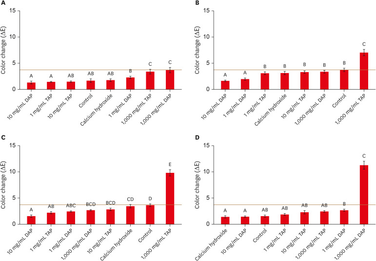
ePub Objectives This study evaluated the effects of low and moderate concentrations of triple antibiotic paste (TAP) and double antibiotic paste (DAP) loaded into a hydrogel system on crown discoloration and explored whether application of an adhesive bonding agent prevented crown discoloration.
Materials and Methods Intact human molars (
n = 160) were horizontally sectioned 1 mm apical to the cementoenamel junction. The crowns were randomized into 8 experimental groups (calcium hydroxide, Ca[OH]2; 1, 10, and 1,000 mg/mL TAP and DAP; and no medicament. The pulp chambers in half of the samples were coated with an adhesive bonding agent before receiving the intracanal medicament. Color changes (ΔE) were detected by spectrophotometry after 1 day, 1 week, and 4 weeks, and after 5,000 thermal cycles, with ΔE = 3.7 as a perceptible threshold. The 1-samplet -test was used to determine the significance of color changes relative to 3.7. Analysis of variance was used to evaluate the effects of treatment, adhesive, and time on color change, and the level of significance wasp < 0.05.Results Ca(OH)2 and 1 and 10 mg/mL DAP did not cause clinically perceivable tooth discoloration. Adhesive agent use significantly decreased tooth discoloration in the 1,000 mg/mL TAP group up to 4 weeks. However, adhesive use did not significantly improve coronal discoloration after thermocycling when 1,000 mg/mL TAP was used.
Conclusions Ca(OH)2 and 1 and 10 mg/mL DAP showed no clinical discoloration. Using an adhesive significantly improved coronal discoloration up to 4 weeks with 1,000 mg/mL TAP.
- 19 View
- 1 Download
- 3 Web of Science

- Conservative approach of a symptomatic carious immature permanent tooth using a tricalcium silicate cement (Biodentine): a case report
- Cyril Villat, Brigitte Grosgogeat, Dominique Seux, Pierre Farge
- Restor Dent Endod 2013;38(4):258-262. Published online November 12, 2013
- DOI: https://doi.org/10.5395/rde.2013.38.4.258
-
 Abstract
 PDF
PDF PubReader
 ePub
ePub The restorative management of deep carious lesions and the preservation of pulp vitality of immature teeth present real challenges for dental practitioners. New tricalcium silicate cements are of interest in the treatment of such cases. This case describes the immediate management and the follow-up of an extensive carious lesion on an immature second right mandibular premolar. Following anesthesia and rubber dam isolation, the carious lesion was removed and a partial pulpotomy was performed. After obtaining hemostasis, the exposed pulp was covered with a tricalcium silicate cement (Biodentine, Septodont) and a glass ionomer cement (Fuji IX extra, GC Corp.) restoration was placed over the tricalcium silicate cement. A review appointment was arranged after seven days, where the tooth was asymptomatic with the patient reporting no pain during the intervening period. At both 3 and 6 mon follow up, it was noted that the tooth was vital, with normal responses to thermal tests. Radiographic examination of the tooth indicated dentin-bridge formation in the pulp chamber and the continuous root formation. This case report demonstrates a fast tissue response both at the pulpal and root dentin level. The use of tricalcium silicate cement should be considered as a conservative intervention in the treatment of symptomatic immature teeth.
- 22 View
- 0 Download

- Influence of post types and sizes on fracture resistance in the immature tooth model
- Jong-Hyun Kim, Sung-Ho Park, Jeong-Won Park, Il-Young Jung
- J Korean Acad Conserv Dent 2010;35(4):257-266. Published online July 31, 2010
- DOI: https://doi.org/10.5395/JKACD.2010.35.4.257
-
 Abstract
 PDF
PDF PubReader
 ePub
ePub The purpose of this study was to determine the effect of post types and sizes on fracture resistance in immature tooth model with various restorative techniques. Bovine incisors were sectioned 8 mm above and 12 mm below the cementoenamel junction to simulate immature tooth model. To compare various post-and-core restorations, canals were restored with gutta-percha and resin core, or reinforced dentin wall with dual-cured resin composite, followed by placement of D.T. LIGHT-POST, ParaPost XT, and various sizes of EverStick Post individually. All of specimens were stored in the distilled water for 72 hours and underwent 6,000 thermal cycles. After simulation of periodontal ligament structure with polyether impression material, compressive load was applied at 45 degrees to the long axis of the specimen until fracture was occurred.
Experimental groups reinforced with post and composite resin were shown significantly higher fracture strength than gutta-percha group without post placement (p < 0.05). Most specimens fractured limited to cervical third of roots. Post types did not influence on fracture resistance and fracture level significantly when cement space was filled with dual-cured resin composite. In addition, no statistically significant differences were seen between customized and standardized glass fiber posts, which cement spaces were filled with resin cement or composite resin individually. Therefore, root reinforcement procedures as above in immature teeth improved fracture resistance regardless of post types and sizes.
- 18 View
- 0 Download


 KACD
KACD
 First
First Prev
Prev


