Search
- Page Path
- HOME > Search
-
Does minimally invasive canal preparation provide higher fracture resistance of endodontically treated teeth? A systematic review of
in vitro studies - Sıla Nur Usta, Emmanuel João Nogueira Leal Silva, Seda Falakaloğlu, Mustafa Gündoğar
- Restor Dent Endod 2023;48(4):e34. Published online October 17, 2023
- DOI: https://doi.org/10.5395/rde.2023.48.e34

-
 Abstract
 PDF
PDF PubReader
 ePub
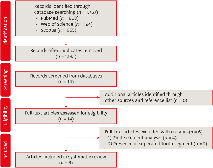
ePub This systematic review aimed to investigate whether minimally invasive root canal preparation ensures higher fracture resistance compared to conventional root canal preparation in endodontically treated teeth (ETT). A comprehensive search strategy was conducted on the “PubMed, Web of Science, and Scopus” databases, alongside reference and hand searches, with language restrictions applied. Two independent reviews selected pertinent laboratory studies that explored the effect of minimally invasive root canal preparation on fracture resistance, in comparison to larger preparation counterparts. The quality of the studies was assessed, and the risk of bias was categorized as low, moderate, or high. The electronic search yielded a total of 1,767 articles. After applying eligibility criteria, 8 studies were included. Given the low methodological quality of these studies and the large variability of fracture resistance values, the impact of reduced apical size and/or taper on the fracture resistance of the ETT can be considered uncertain. This systematic review could not reveal sufficient evidence regarding the effect of minimally invasive preparation on increasing fracture resistance of ETT, primarily due to the inherent limitations of the studies and the moderate risk of bias.
-
Citations
Citations to this article as recorded by- Impact of conservative versus conventional instrumentation on the release of inflammatory mediators and post‐operative pain in mandibular molars with asymptomatic irreversible pulpitis: A randomized clinical trial
Sıla Nur Usta, Ana Arias, Emre Avcı, Emmanuel João Nogueira Leal Silva
International Endodontic Journal.2025; 58(6): 862. CrossRef - Mapping risk of bias criteria in systematic reviews of in vitro endodontic studies: an umbrella review
Rafaella Rodrigues da Gama, Lucas Peixoto de Araújo, Evandro Piva, Leandro Perello Duro, Adriana Fernandes da Silva, Wellington Luiz de Oliveira da Rosa
Evidence-Based Dentistry.2025; 26(4): 179. CrossRef - Micro‐computed tomography evaluation of minimally invasive root canal preparation in 3D‐printed C‐shaped canal
Nutcha Supavititpattana, Siriwan Suebnukarn, Panupat Phumpatrakom, Kamon Budsaba
Australian Endodontic Journal.2024; 50(3): 621. CrossRef - Ex vivo investigation on the effect of minimally invasive endodontic treatment on vertical root fracture resistance and crack formation
Andreas Rathke, Henry Frehse, Maria Bechtold
Scientific Reports.2024;[Epub] CrossRef
- Impact of conservative versus conventional instrumentation on the release of inflammatory mediators and post‐operative pain in mandibular molars with asymptomatic irreversible pulpitis: A randomized clinical trial
- 4,241 View
- 120 Download
- 4 Web of Science
- 4 Crossref

- Cutting efficiency of apical preparation using ultrasonic tips with microprojections: confocal laser scanning microscopy study
- Sang-Won Kwak, Young-Mi Moon, Yeon-Jee Yoo, Seung-Ho Baek, WooCheol Lee, Hyeon-Cheol Kim
- Restor Dent Endod 2014;39(4):276-281. Published online July 22, 2014
- DOI: https://doi.org/10.5395/rde.2014.39.4.276
-
 Abstract
 PDF
PDF PubReader
 ePub
ePub Objectives The purpose of this study was to compare the cutting efficiency of a newly developed microprojection tip and a diamond-coated tip under two different engine powers.
Materials and Methods The apical 3-mm of each root was resected, and root-end preparation was performed with upward and downward pressure using one of the ultrasonic tips, KIS-1D (Obtura Spartan) or JT-5B (B&L Biotech Ltd.). The ultrasonic engine was set to power-1 or -4. Forty teeth were randomly divided into four groups: K1 (KIS-1D / Power-1), J1 (JT-5B / Power-1), K4 (KIS-1D / Power-4), and J4 (JT-5B / Power-4). The total time required for root-end preparation was recorded. All teeth were resected and the apical parts were evaluated for the number and length of cracks using a confocal scanning micrscope. The size of the root-end cavity and the width of the remaining dentin were recorded. The data were statistically analyzed using two-way analysis of variance and a Mann-Whitney test.
Results There was no significant difference in the time required between the instrument groups, but the power-4 groups showed reduced preparation time for both instrument groups (
p < 0.05). The K4 and J4 groups with a power-4 showed a significantly higher crack formation and a longer crack irrespective of the instruments. There was no significant difference in the remaining dentin thickness or any of the parameters after preparation.Conclusions Ultrasonic tips with microprojections would be an option to substitute for the conventional ultrasonic tips with a diamond coating with the same clinical efficiency.
-
Citations
Citations to this article as recorded by- Effectiveness of Sectioning Method and Filling Materials on Roughness and Cell Attachments in Root Resection Procedure
Tarek Ashi, Naji Kharouf, Olivier Etienne, Bérangère Cournault, Pierre Klienkoff, Varvara Gribova, Youssef Haikel
European Journal of Dentistry.2025; 19(01): 240. CrossRef - Questioning the spot light on Hi-tech endodontics
Jojo Kottoor, Denzil Albuquerque
Restorative Dentistry & Endodontics.2016; 41(1): 80. CrossRef
- Effectiveness of Sectioning Method and Filling Materials on Roughness and Cell Attachments in Root Resection Procedure
- 1,390 View
- 6 Download
- 2 Crossref

- Evaluation of canal preparation for apical sealing with various Ni-Ti rotary instruments
- Yooseok Shin, Su-Jung Shin, Minju Song, Euiseong Kim
- J Korean Acad Conserv Dent 2011;36(4):300-305. Published online July 31, 2011
- DOI: https://doi.org/10.5395/JKACD.2011.36.4.300
-
 Abstract
 PDF
PDF PubReader
 ePub
ePub Objectives The aim of this study was to evaluate the various NiTi rotary instruments regarding their ability to provide a circular apical preparation.
Materials and Methods 50 single canal roots were selected, cut at the cementodentinal junction and the coronal 1/3 of the canals was flared using Gates Glidden burs. Samples were randomly divided into 5 experimental groups of 10 each. In group I, GT files, Profile 04 and Quantec #9 and #10 files were used. In Group II Lightspeed was used instead of Quantec. In Group III, Orifice shaper, Profile .06 series and Lightspeed were used. In Group IV, Quantec #9 and #10 files were used instead of Lightspeed. In Group V, the GT file and the Profile .04 series were used to prepare the entire canal length. All tooth samples were cut at 1 mm, 3 mm and 5 mm from the apex and were examined under the microscope.
Results Groups II and III (Lightspeed) showed a more circular preparation in the apical 1mm samples than the groups that used Quantec (Group I & IV) or GT files and Profile .04 series.(Group V)(
p < 0.05) There was no significant difference statistically among the apical 3, 5 mm samples. In 5 mm samples, most of the samples showed complete circularity and none of them showed irregular shape.Conclusions Lightspeed showed circular preparation at apical 1 mm more frequently than other instruments used in this study. However only 35% of samples showed circularity even in the Lightspeed Group which were enlarged 3 ISO size from the initial apical binding file (IAF) size. So it must be considered that enlarging 3 ISO size isn't enough to make round preparation.
- 719 View
- 5 Download


 KACD
KACD

 First
First Prev
Prev


