The prevalence of radix molaris in the mandibular first molars of a Saudi subpopulation based on cone-beam computed tomography
Article information
Abstract
Objectives
The purpose of this study was to determine the incidence of radix molaris (RM) (entomolaris and paramolaris) in the mandibular first permanent molars of a sample Saudi Arabian subpopulation using cone-beam computed tomography (CBCT).
Materials and Methods
A total of 884 CBCT images of 427 male and 457 female Saudi citizens (age 16 to 70 years) were collected from the radiology department archives of 4 dental centers. A total of 450 CBCT images of 741 mature mandibular first molars that met the inclusion criteria were reviewed. The images were viewed at high resolution by 3 examiners and were analyzed with Planmeca Romexis software (version 5.2).
Results
Thirty-three (4.5%) mandibular first permanent molars had RM, mostly on the distal side. The incidence of radix entomolaris (EM) was 4.3%, while that of radix paramolaris was 0.3%. The RM roots had one canal and occurred more unilaterally. No significant difference in root configuration was found between males and females (p > 0.05). Types I and III EM root canal configurations were most common, while type B was the only RP configuration observed.
Conclusions
The incidence of RM in the mandibular first molars of this Saudi subpopulation was 4.5%. Identification of the supernumerary root can avoid missing the canal associated with the root during root canal treatment.
INTRODUCTION
In-depth knowledge of the internal anatomy of the root canal system and its variations is important for successful root canal treatment [1]. Preoperative radiographic imaging provides much-needed information about root canal morphology, including the No. of roots and canals. This allows the clinician to assess the correct course of endodontic treatment and increase the chance of a successful outcome [23]. The mandibular first molars usually have 3 or 4 canals and 2 roots; however, the No. of root canals and roots may vary. The presence of a supernumerary root in the mandibular first molar is called radix molaris (RM). The supernumerary root is usually located distolingually (radix entomolaris [EM]) with a global prevalence of 0.7%–33.1% [4567891011121314151617181920] (Table 1). When the extra root is located on the mesiobuccal side, it is called radix paramolaris (PM). Several studies have demonstrated differences in root canal configurations such as shape and the No. of roots and canals among different populations [212223]. The reported prevalence of 3-rooted mandibular first molars in Saudi Arabia ranges from 2.3%–6% based on visual examinations of extracted teeth [724] and conventional radiographs in clinical cases of endodontically-treated teeth [10].
Conventional radiographs are the most common diagnostic tool used to evaluate the morphology of mandibular roots [25]. Although radiographs can demonstrate the main morphological features of the tooth, the complexities and details of the root canal anatomy can be harder to visualize due to the use of 2-dimensional (2D) images to view a 3-dimensional (3D) object [26]. Cone-beam computed tomography (CBCT) has excellent resolution and can provide better visualization of the external and internal morphologies of the tooth and its root canal system compared to conventional and digital radiographs [2728]. Therefore, CBCT can be especially helpful in clinical cases regarding root canal morphology. The purpose of this study was to determine the incidence of RM using CBCT of the mandibular first permanent molars in a sample Saudi Arabian subpopulation.
MATERIALS AND METHODS
This descriptive-analytic study was approved by the Ethical Committee of the Research Center of Riyadh Elm University College of Dentistry (RC/IRB/2018/1086). The study was conducted in compliance with the Helsinki Declaration and Guidelines for Good Clinical Practice.
Sample collection
Data were collected from archived CBCT images taken from 2014–2018 at the radiology departments of 4 dental centers: the College of Dentistry of Riyadh Elm University, King Saud University, Prince Sattam Bin Abdulaziz University, and Uranus Dental Center. A total of 884 CBCT images of 427 male and 457 female Saudi citizens were reviewed (Table 2). The CBCT images were captured using 3 different CBCT machines: the Promax 3D Max (Planmeca, Helsinki, Finland) (90 kVp, 10 mA, 10–15 seconds) and the Galileos Comfort (Sirona Dental Systems GmbH, Bensheim, Germany) (85 kVp, 7 mA, 14 seconds) using an isotropic voxel size of 0.2–0.4 mm, and the CS9300 (Carestream Dental LLC, Atlanta, GA, USA) (90 kVp, 10–15 mA, 15 seconds) using an isotropic voxel size of 0.9 mm. Images of mandibular first molars that met the following inclusion criteria were chosen for evaluation: 1) fully erupted first mandibular permanent molar; 2) no periapical lesions, resorption, or canal calcification; 3) no root canal fillings, cemented posts, or coronal restoration; 4) bilateral or unilateral location; and 5) mature (closed) root apices. A total of 450 CBCT images met the inclusion criteria.
Evaluation of CBCT images
Three examiners evaluated the CBCT images, 1 postgraduate (PG) endodontic resident in the last year (referred to as ‘examiner’) and 2 certified oral radiologists (referred to as ‘radiologist’). Prior to the evaluation of the CBCT images, the PG resident was trained in the manipulation of CBCT images, locating the supernumerary root, and assessing the root canal configuration. The CBCT images were analyzed with Planmeca Romexis software (version 5.2; Planmeca). Images were viewed on a 32 in the LCD monitor (Hewlett-Packard, Palo Alto, CA, USA) at a resolution of 1,280 × 1,024 pixels. The classification systems of Carlsen and Alexandersen [29] and Song et al. [30] were used for the categorization of the root configurations. The image evaluations were done under sufficient magnification and contrast to ensure optimal visualization. Extra roots, when present, were classified as either entomolaris or paramolaris.
Statistical analysis
All statistical analyses were performed using SPSS software (version 20; IBM Corp., Armonk, NY, USA). To establish inter- and intra-examiner reliability, ten cases were selected to be read by the examiner and radiologists and were measured using Cohen's kappa coefficient. The Kappa agreement test of the reading between the examiners was 97%, which considered the solid agreement and statistically significant (p ≤ 0.05). Continuous data were evaluated using descriptive statistics. The χ2 test was used to assess the differences in root configuration classification between males and females.
RESULTS
Root morphology
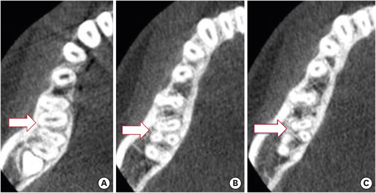
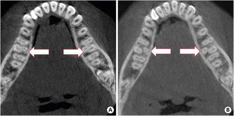
Thirty-three (4.5%) mandibular first permanent molars had supernumerary roots, mostly on the distal side (Figure 1, Table 3). The incidence of EM was 4.2%, while that of PM was 0.3% (Table 4). The RM roots predominantly had one canal and most often occurred unilaterally. The bilateral occurrence was 1.3% (Figure 2). There was no significant difference in root configuration between males and females (p > 0.05).

(A) Cone-beam computed tomography (CBCT) images of mandibular first molar showing 4 canals (arrow). (B) CBCT showing disto-buccal root with one canal (arrow). (C) Separated distal roots with one canal each (arrow).

Number and percentages of patients with entomolaris (distolingual root) and paramolaris (mesiobuccal root) in mandibular first molars according to sex and jaw side (n = 741)
DISCUSSION
The prevalence of RM in the current study was 4.5%, which is close to those values reported by Rodrigues et al. [18] and Rahimi et al. [19] and far less than those reported by Zhang et al. [15], Gupta et al. [20], and Wang et al. [31]. The occurrence of RM in both males and females in the current study was predominantly unilateral (n = 23, 3.1%). This value is far lower than in a previous study by Quackenbush [32], which reported RM occurring unilaterally in approximately 40% of all cases.
Previous studies have used extracted teeth to determine the frequency of RM. This technique might underestimate the true frequency because small supernumerary roots might easily fracture during extraction [56213334]. Periapical radiographs from different angles can also be done to identify supernumerary roots. An angulated main beam is usually used to avoid superimposition of the larger distobuccal root [33]. Although this technique may accurately capture the tooth's main morphological features, the complexities and details of the root canal's anatomy cannot be shown due to the nature of using 2D images to visualize a 3D object [26]. The identification of additional supernumerary roots in the distal and/or mesial aspect of the mandibular first molar is considered important for the long-term outcome of the endodontically-treated tooth.
The present study used CBCT to determine the occurrence of RM in the permanent mandibular molars among a Saudi subpopulation. Patel et al. [35] reported that the root morphology and the No. of root canals and their convergence or divergence from each other are better visualized using 3D imaging techniques. The use of CBCT provides the clinician with the ability to observe an area in 3 different planes (sagittal, coronal, and axial), which has been reported to eliminate the superimposition of anatomic structures [3637]. In addition, Neelakantan et al. [37] reported that CBCT and peripheral quantitative computed tomography were as accurate as the modified canal staining and tooth clearing technique for the study of canal morphology. Furthermore, Matherne et al. [27] stated that CBCT could identify a greater No. of root canal systems than digital radiographs. Compared to conventional medical computed tomography, CBCT allows less scan time, a lower radiation dose, and higher resolution imaging [37].
The incidence of RM in mandibular first permanent molars has been reported as being associated with certain ethnic groups. RM has been found more frequently in Asian populations than in other racial groups, especially in China and Korea (27%–33%) [4141538]. In the Asian race, it is considered to be a normal morphological variant. In contrast, the incidence of RM has not been reported in Turkish [23], Spanish [39], Pakistani [40], or Ugandan populations [41]. Other studies using extracted teeth or periapical radiographs reported similar findings, including those of Zaatar et al. [8] in a Kuwaiti population, Sperber and Moreau [9] in a Senegalese population, Ahmed et al. [11] in a Sudanese population, Al-Qudah and Awawdeh [13] in a Jordanian population, and Mukhaimer and Azizi [17] in a Palestinian population.
In comparison to previous studies evaluating RM in a Saudi population, the findings of the current study are higher than those of Younes et al. [7] using extracted teeth (2.3%), and lower than the clinical and radiographic evaluation by Al-Nazhan [10] (5.97%) and extraction evaluation by Bahammam and Bahammam [24] (6.0%). Teeth that were clinically and radiographically evaluated by Al-Nazhan [10] were difficult cases referred to the endodontic clinic. Similar findings were reported by Song et al. [30], who reported a frequency of 24.5% when using computed tomography compared to 33.1% when using periapical radiographs [14]. They concluded that the relationship of the distolingual roots between the first and second molars could be explained by the developmental theory [42]. They further added that the occurrence of distolingual roots might be regarded as a characteristic feature of the field in which the first permanent molar is the key tooth.
Different classification systems have been used to describe the morphological features of supernumerary roots in mandibular molars [142943]. The morphology of RM in the present study was classified according to Carlsen and Alexandersen [29] and Song et al. [30]. In the case of a mesiobuccal root (PM), Carlsen and Alexandersen [29] examined 203 permanent molars and found 5 cases of type A and none of type B. In the current study, we found only 2 cases of type B and none of type A. This difference could be related to ethnicity. Regarding the distolingual root (EM), Song et al. [30] examined computed tomography images of first mandibular molars and divided the morphologic features of the supernumerary root into 5 types according to the pattern of their morphology. In the current study, we predominantly saw Types I and III. This is different from the findings of Song et al. [30] who reported type II as the most predominant, and this could be related to ethnicity.
The etiology behind the formation of supernumerary roots in mandibular molars is unclear. Their formation could be related to external factors during odontogenesis or to an atavistic or polygenetic gene [44]. Kim et al. [45] reported that the crowns of first permanent molars with distolingual extra-roots had significantly larger intercuspal distances between the distobuccal and distolingual cusp tips and a larger distal buccolingual width than those without. Furthermore, the distance from the EM (distolingual root) apex to the outer surface of the buccal cortical bone was calculated by Kim et al. [46] and found it to be within 12.09 mm. They concluded that the knowledge of the anatomic and morphologic of the mandibular first molar could be useful in endodontic treatment planning. In addition, the outline of the pulp chamber in molars with 3 root canals is triangular, while molars with 4 canals where the extra root (RM) might be present have a rectangular or rhomboidal chamber [9]. This should be considered during root canal treatment to avoid missing the canal of the extra root.
The presence of EM or PM has important clinical implications in endodontic therapy. Identification of these supernumerary roots can avoid missing the canal associated with the root during root canal treatment. EM root is often located in the same plane of the PM root, and superimposition of both roots can appear on conventional radiographs, leading in an inaccurate diagnosis. To reveal the EM, a second radiograph from a more mesial or distal angle or using CBCT should be taken. A CBCT image will show the exact position of the supernumerary root and helps in tracking its curvature in order to avoid procedural errors during root canal treatment.
CONCLUSIONS
Within the limitations of this study, we found that the incidence of RM in the mandibular first molars of a Saudi subpopulation was 4.5%. In addition, we found that CBCT is an accurate, reliable, non-invasive, and practical technique for identifying RM in mandibular first molars.
Notes
Conflict of Interest: No potential conflict of interest relevant to this article was reported.
Author Contributions:
Conceptualization: AL-Alawi H, Aldosimani MA, Zahid MN.
Data curation: AL-Alawi H, Shihabi GN.
Formal analysis: Al-Nazhan S, AL-Alawi H.
Investigation: AL-Alawi H, Al-Nazhan S, Al-Maflehi N, Aldosimani MA, Zahid MN, Shihabi GN.
Methodology: Al-Nazhan S.
Project administration: AL-Alawi H.
Resources: AL-Alawi H, Aldosimani MA, Zahid MN.
Software: Al-Maflehi N.
Supervision: Al-Nazhan S, AL-Alawi H.
Validation: Al-Nazhan S, AL-Alawi H, Al-Maflehi N.
Visualization: Al-Nazhan S, AL-Alawi H, Aldosimani MA, Zahid MN, Shihabi GN.
Writing - original draft: Al-Nazhan S.
Writing - review & editing: Al-Nazhan S.


